Impact of scaffold rigidity on the design and evolution of an artificial Diels-Alderase.
Preiswerk, N., Beck, T., Schulz, J.D., Milovnik, P., Mayer, C., Siegel, J.B., Baker, D., Hilvert, D.(2014) Proc Natl Acad Sci U S A 111: 8013-8018
- PubMed: 24847076 Search on PubMedSearch on PubMed Central
- DOI: https://doi.org/10.1073/pnas.1401073111
- Primary Citation Related Structures:
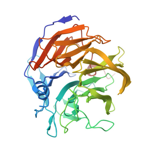
4O5S, 4O5T - PubMed Abstract:
By combining targeted mutagenesis, computational refinement, and directed evolution, a modestly active, computationally designed Diels-Alderase was converted into the most proficient biocatalyst for [4+2] cycloadditions known. The high stereoselectivity and minimal product inhibition of the evolved enzyme enabled preparative scale synthesis of a single product diastereomer. X-ray crystallography of the enzyme-product complex shows that the molecular changes introduced over the course of optimization, including addition of a lid structure, gradually reshaped the pocket for more effective substrate preorganization and transition state stabilization. The good overall agreement between the experimental structure and the original design model with respect to the orientations of both the bound product and the catalytic side chains contrasts with other computationally designed enzymes. Because design accuracy appears to correlate with scaffold rigidity, improved control over backbone conformation will likely be the key to future efforts to design more efficient enzymes for diverse chemical reactions.
- Laboratory of Organic Chemistry, Eidgenössische Technische Hochschule Zürich, 8093 Zurich, Switzerland;
Organizational Affiliation: