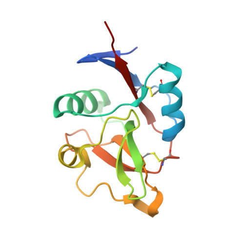
4N36

Structure of langerin CRD I313 D288 complexed with Me-GlcNAc


Experimental Data Snapshot

  • Method: X-RAY DIFFRACTION
  • Resolution: 1.85 Å
  • R-Value Free: 0.214 
  • R-Value Work: 0.170 
  • R-Value Observed: 0.172 

Starting Model: experimental
View more details

wwPDB Validation   3D Report Full Report


Ligand Structure Quality Assessment 


This is version 1.5 of the entry. See complete history


Literature

Common polymorphisms in human langerin change specificity for glycan ligands.

Feinberg, H.Rowntree, T.J.Tan, S.L.Drickamer, K.Weis, W.I.Taylor, M.E.

(2013) J Biol Chem 288: 36762-36771

  • DOI: https://doi.org/10.1074/jbc.M113.528000
  • Primary Citation of Related Structures:  
    4N32, 4N33, 4N34, 4N35, 4N36, 4N37, 4N38

  • PubMed Abstract: 

    Langerin, a C-type lectin on Langerhans cells, mediates carbohydrate-dependent uptake of pathogens in the first step of antigen presentation to the adaptive immune system. Langerin binds a diverse range of carbohydrates including high mannose structures, fucosylated blood group antigens, and glycans with terminal 6-sulfated galactose. Mutagenesis and quantitative binding assays indicate that salt bridges between the sulfate group and two lysine residues compensate for the nonoptimal binding of galactose at the primary Ca(2+) site. A commonly occurring single nucleotide polymorphism (SNP) in human langerin results in change of one of these lysine residues, Lys-313, to isoleucine. Glycan array screening reveals that this amino acid change abolishes binding to oligosaccharides with terminal 6SO4-Gal and enhances binding to oligosaccharides with terminal GlcNAc residues. Structural analysis shows that enhanced binding to GlcNAc may result from Ile-313 packing against the N-acetyl group. The K313I polymorphism is tightly linked to another SNP that results in the change N288D, which reduces affinity for glycan ligands by destabilizing the Ca(2+)-binding site. Langerin with Asp-288 and Ile-313 shows no binding to 6SO4-Gal-terminated glycans and increased binding to GlcNAc-terminated structures, but overall decreased binding to glycans. Altered langerin function in individuals with the linked N288D and K313I polymorphisms may affect susceptibility to infection by microorganisms.


  • Organizational Affiliation

    From the Department of Life Sciences, Imperial College, London SW7 2AZ, United Kingdom and.


Macromolecules
Find similar proteins by:  (by identity cutoff)  |  3D Structure
Entity ID: 1
MoleculeChains Sequence LengthOrganismDetailsImage
C-type lectin domain family 4 member K
A, B, C, D
136Homo sapiensMutation(s): 3 
Gene Names: CD207CLEC4K
UniProt & NIH Common Fund Data Resources
Find proteins for Q9UJ71 (Homo sapiens)
Explore Q9UJ71 
Go to UniProtKB:  Q9UJ71
PHAROS:  Q9UJ71
GTEx:  ENSG00000116031 
Entity Groups  
Sequence Clusters30% Identity50% Identity70% Identity90% Identity95% Identity100% Identity
UniProt GroupQ9UJ71
Sequence Annotations
Expand
  • Reference Sequence
Small Molecules
Ligands 3 Unique
IDChains Name / Formula / InChI Key2D Diagram3D Interactions
2F8
Query on 2F8

Download Ideal Coordinates CCD File 
E [auth A],
G [auth B],
I [auth C],
K [auth D]
methyl 2-acetamido-2-deoxy-alpha-D-glucopyranoside
C9 H17 N O6
ZEVOCXOZYFLVKN-OKNNCHMLSA-N
CA
Query on CA

Download Ideal Coordinates CCD File 
F [auth A],
H [auth B],
J [auth C],
L [auth D]
CALCIUM ION
Ca
BHPQYMZQTOCNFJ-UHFFFAOYSA-N
MG
Query on MG

Download Ideal Coordinates CCD File 
M [auth D]MAGNESIUM ION
Mg
JLVVSXFLKOJNIY-UHFFFAOYSA-N
Experimental Data & Validation

Experimental Data

  • Method: X-RAY DIFFRACTION
  • Resolution: 1.85 Å
  • R-Value Free: 0.214 
  • R-Value Work: 0.170 
  • R-Value Observed: 0.172 
  • Space Group: P 42
Unit Cell:
Length ( Å )Angle ( ˚ )
a = 79.55α = 90
b = 79.55β = 90
c = 89.88γ = 90
Software Package:
Software NamePurpose
SCALAdata scaling
PHENIXrefinement
PDB_EXTRACTdata extraction
Blu-Icedata collection
XDSdata reduction
PHENIXphasing

Structure Validation

View Full Validation Report


Ligand Structure Quality Assessment 


Entry History 

Deposition Data

Revision History  (Full details and data files)

  • Version 1.0: 2013-11-20
    Type: Initial release
  • Version 1.1: 2013-12-04
    Changes: Database references
  • Version 1.2: 2014-01-22
    Changes: Database references
  • Version 1.3: 2017-11-15
    Changes: Refinement description
  • Version 1.4: 2020-07-29
    Type: Remediation
    Reason: Carbohydrate remediation
    Changes: Data collection, Database references, Derived calculations, Structure summary
  • Version 1.5: 2023-09-20
    Changes: Data collection, Database references, Refinement description, Structure summary