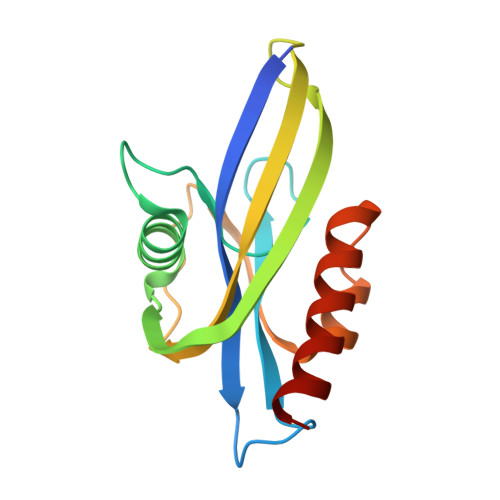
4MPO

1.90 A resolution structure of CT771 from Chlamydia trachomatis Bound to Hydrolyzed Ap4A Products


Experimental Data Snapshot

  • Method: X-RAY DIFFRACTION
  • Resolution: 1.90 Å
  • R-Value Free: 0.250 
  • R-Value Work: 0.203 
  • R-Value Observed: 0.206 

Starting Model: experimental
View more details

wwPDB Validation   3D Report Full Report


Ligand Structure Quality Assessment 


This is version 1.3 of the entry. See complete history


Literature

Chlamydia trachomatis CT771 (nudH) Is an Asymmetric Ap4A Hydrolase.

Barta, M.L.Lovell, S.Sinclair, A.N.Battaile, K.P.Hefty, P.S.

(2014) Biochemistry 53: 214-224

  • DOI: https://doi.org/10.1021/bi401473e
  • Primary Citation of Related Structures:  
    4ILQ, 4MPO

  • PubMed Abstract: 

    Asymmetric diadenosine 5',5‴-P(1),P(4)-tetraphosphate (Ap4A) hydrolases are members of the Nudix superfamily that asymmetrically cleave the metabolite Ap4A into ATP and AMP while facilitating homeostasis. The obligate intracellular mammalian pathogen Chlamydia trachomatis possesses a single Nudix family protein, CT771. As pathogens that rely on a host for replication and dissemination typically have one or zero Nudix family proteins, this suggests that CT771 could be critical for chlamydial biology and pathogenesis. We identified orthologues to CT771 within environmental Chlamydiales that share active site residues suggesting a common function. Crystal structures of both apo- and ligand-bound CT771 were determined to 2.6 Å and 1.9 Å resolution, respectively. The structure of CT771 shows a αβα-sandwich motif with many conserved elements lining the putative Nudix active site. Numerous aspects of the ligand-bound CT771 structure mirror those observed in the ligand-bound structure of the Ap4A hydrolase from Caenorhabditis elegans. These structures represent only the second Ap4A hydrolase enzyme member determined from eubacteria and suggest that mammalian and bacterial Ap4A hydrolases might be more similar than previously thought. The aforementioned structural similarities, in tandem with molecular docking, guided the enzymatic characterization of CT771. Together, these studies provide the molecular details for substrate binding and specificity, supporting the analysis that CT771 is an Ap4A hydrolase (nudH).


  • Organizational Affiliation

    Department of Molecular Biosciences, University of Kansas , Lawrence, Kansas 66045, United States.


Macromolecules
Find similar proteins by:  (by identity cutoff)  |  3D Structure
Entity ID: 1
MoleculeChains Sequence LengthOrganismDetailsImage
CT771
A, B, C, D, E
A, B, C, D, E, F, G, H
153Chlamydia trachomatis L2/434/BuMutation(s): 0 
Gene Names: CTL0140
EC: 3
UniProt
Find proteins for A0A0H3MCJ9 (Chlamydia trachomatis serovar L2 (strain ATCC VR-902B / DSM 19102 / 434/Bu))
Explore A0A0H3MCJ9 
Go to UniProtKB:  A0A0H3MCJ9
Entity Groups  
Sequence Clusters30% Identity50% Identity70% Identity90% Identity95% Identity100% Identity
UniProt GroupA0A0H3MCJ9
Sequence Annotations
Expand
  • Reference Sequence
Small Molecules
Ligands 4 Unique
IDChains Name / Formula / InChI Key2D Diagram3D Interactions
AMP
Query on AMP

Download Ideal Coordinates CCD File 
BA [auth D]
J [auth A]
JA [auth E]
NA [auth F]
P [auth B]
BA [auth D],
J [auth A],
JA [auth E],
NA [auth F],
P [auth B],
SA [auth G],
X [auth C],
XA [auth H]
ADENOSINE MONOPHOSPHATE
C10 H14 N5 O7 P
UDMBCSSLTHHNCD-KQYNXXCUSA-N
PO4
Query on PO4

Download Ideal Coordinates CCD File 
CA [auth D]
IA [auth E]
K [auth A]
OA [auth F]
Q [auth B]
CA [auth D],
IA [auth E],
K [auth A],
OA [auth F],
Q [auth B],
TA [auth G],
W [auth C],
YA [auth H]
PHOSPHATE ION
O4 P
NBIIXXVUZAFLBC-UHFFFAOYSA-K
CL
Query on CL

Download Ideal Coordinates CCD File 
AA [auth D],
DB [auth H],
HA [auth E],
I [auth A],
O [auth B]
CHLORIDE ION
Cl
VEXZGXHMUGYJMC-UHFFFAOYSA-M
MG
Query on MG

Download Ideal Coordinates CCD File 
AB [auth H]
BB [auth H]
CB [auth H]
DA [auth D]
EA [auth D]
AB [auth H],
BB [auth H],
CB [auth H],
DA [auth D],
EA [auth D],
FA [auth D],
GA [auth E],
KA [auth E],
L [auth A],
LA [auth E],
M [auth A],
MA [auth E],
N [auth A],
PA [auth F],
QA [auth F],
R [auth B],
RA [auth F],
S [auth B],
T [auth B],
U [auth C],
UA [auth G],
V [auth C],
VA [auth G],
WA [auth G],
Y [auth C],
Z [auth C],
ZA [auth H]
MAGNESIUM ION
Mg
JLVVSXFLKOJNIY-UHFFFAOYSA-N
Experimental Data & Validation

Experimental Data

  • Method: X-RAY DIFFRACTION
  • Resolution: 1.90 Å
  • R-Value Free: 0.250 
  • R-Value Work: 0.203 
  • R-Value Observed: 0.206 
  • Space Group: C 1 2 1
Unit Cell:
Length ( Å )Angle ( ˚ )
a = 182.528α = 90
b = 102.813β = 94.03
c = 65.363γ = 90
Software Package:
Software NamePurpose
Aimlessdata scaling
PHENIXrefinement
PDB_EXTRACTdata extraction
JDirectordata collection
XDSdata reduction
PHASERphasing

Structure Validation

View Full Validation Report


Ligand Structure Quality Assessment 


Entry History 

Deposition Data

Revision History  (Full details and data files)

  • Version 1.0: 2014-01-22
    Type: Initial release
  • Version 1.1: 2014-01-29
    Changes: Database references
  • Version 1.2: 2017-11-15
    Changes: Refinement description
  • Version 1.3: 2023-09-20
    Changes: Data collection, Database references, Derived calculations, Refinement description