A phosphatidylinositol transfer protein integrates phosphoinositide signaling with lipid droplet metabolism to regulate a developmental program of nutrient stress-induced membrane biogenesis.
Ren, J., Pei-Chen Lin, C., Pathak, M.C., Temple, B.R., Nile, A.H., Mousley, C.J., Duncan, M.C., Eckert, D.M., Leiker, T.J., Ivanova, P.T., Myers, D.S., Murphy, R.C., Brown, H.A., Verdaasdonk, J., Bloom, K.S., Ortlund, E.A., Neiman, A.M., Bankaitis, V.A.(2014) Mol Biol Cell 25: 712-727
- PubMed: 24403601 Search on PubMedSearch on PubMed Central
- DOI: https://doi.org/10.1091/mbc.E13-11-0634
- Primary Citation Related Structures:
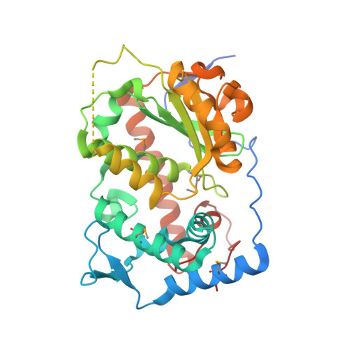
4M8Z - PubMed Abstract:
Lipid droplet (LD) utilization is an important cellular activity that regulates energy balance and release of lipid second messengers. Because fatty acids exhibit both beneficial and toxic properties, their release from LDs must be controlled. Here we demonstrate that yeast Sfh3, an unusual Sec14-like phosphatidylinositol transfer protein, is an LD-associated protein that inhibits lipid mobilization from these particles. We further document a complex biochemical diversification of LDs during sporulation in which Sfh3 and select other LD proteins redistribute into discrete LD subpopulations. The data show that Sfh3 modulates the efficiency with which a neutral lipid hydrolase-rich LD subclass is consumed during biogenesis of specialized membrane envelopes that package replicated haploid meiotic genomes. These results present novel insights into the interface between phosphoinositide signaling and developmental regulation of LD metabolism and unveil meiosis-specific aspects of Sfh3 (and phosphoinositide) biology that are invisible to contemporary haploid-centric cell biological, proteomic, and functional genomics approaches.
- Lineberger Comprehensive Cancer Center, School of Medicine, University of North Carolina at Chapel Hill, Chapel Hill, NC 27599-7090 Department of Molecular and Cellular Medicine, Department of Biochemistry and Biophysics, Texas A&M University, College Station, TX 77843-1114 Department of Biochemistry and Cell Biology, Stony Brook University, Stony Brook, NY 11794-5215 Department of Biochemistry, Emory University School of Medicine, Atlanta, GA 30322-4250 R. L. Juliano Structural Bioinformatics Core, University of North Carolina at Chapel Hill, Chapel Hill, NC 27599-7260 Department of Biology, University of North Carolina at Chapel Hill, Chapel Hill, NC 27599-3280 Department of Biochemistry, University of Utah School of Medicine, Salt Lake City, UT 84112-5650 Department of Pharmacology, University of Colorado Health Sciences Center, Denver, CO 80045-0511 Department of Pharmacology, Vanderbilt University School of Medicine, Nashville, TN 37232-6600.
Organizational Affiliation: