4LW5

Crystal structure of all-trans green fluorescent protein


Experimental Data Snapshot

  • Method: X-RAY DIFFRACTION
  • Resolution: 2.55 Å
  • R-Value Free: 0.281 
  • R-Value Work: 0.218 

wwPDB Validation   3D Report Full Report


This is version 1.3 of the entry. See complete history


Literature

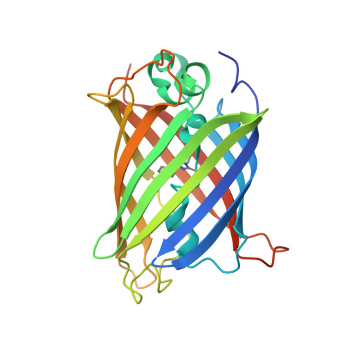
Green-lighting green fluorescent protein: Faster and more efficient folding by eliminating a cis-trans peptide isomerization event.

Rosenman, D.J.Huang, Y.M.Xia, K.Fraser, K.Jones, V.E.Lamberson, C.M.Van Roey, P.Colon, W.Bystroff, C.

(2014) Protein Sci 23: 400-410

  • DOI: https://doi.org/10.1002/pro.2421
  • Primary Citation of Related Structures:  
    4LW5

  • PubMed Abstract: 

    Wild-type green fluorescent protein (GFP) folds on a time scale of minutes. The slow step in folding is a cis-trans peptide bond isomerization. The only conserved cis-peptide bond in the native GFP structure, at P89, was remodeled by the insertion of two residues, followed by iterative energy minimization and side chain design. The engineered GFP was synthesized and found to fold faster and more efficiently than its template protein, recovering 50% more of its fluorescence upon refolding. The slow phase of folding is faster and smaller in amplitude, and hysteresis in refolding has been eliminated. The elimination of a previously reported kinetically trapped state in refolding suggests that X-P89 is trans in the trapped state. A 2.55 Å resolution crystal structure revealed that the new variant contains only trans-peptide bonds, as designed. This is the first instance of a computationally remodeled fluorescent protein that folds faster and more efficiently than wild type.


  • Organizational Affiliation

    Rensselaer Polytechnic Institute, Biological Sciences, 110 8th St., Troy, New York, 12180.


Macromolecules
Find similar proteins by:  (by identity cutoff)  |  3D Structure
Entity ID: 1
MoleculeChains Sequence LengthOrganismDetailsImage
Green fluorescent protein
A, B, C, D, E
239Aequorea victoriaMutation(s): 23 
Gene Names: GFP
UniProt
Find proteins for P42212 (Aequorea victoria)
Explore P42212 
Go to UniProtKB:  P42212
Entity Groups  
Sequence Clusters30% Identity50% Identity70% Identity90% Identity95% Identity100% Identity
UniProt GroupP42212
Sequence Annotations
Expand
  • Reference Sequence
Small Molecules
Modified Residues  1 Unique
IDChains TypeFormula2D DiagramParent
CRO
Query on CRO
A, B, C, D, E
L-PEPTIDE LINKINGC15 H17 N3 O5THR, TYR, GLY
Experimental Data & Validation

Experimental Data

  • Method: X-RAY DIFFRACTION
  • Resolution: 2.55 Å
  • R-Value Free: 0.281 
  • R-Value Work: 0.218 
  • Space Group: P 21 21 21
Unit Cell:
Length ( Å )Angle ( ˚ )
a = 95.557α = 90
b = 110.939β = 90
c = 114.706γ = 90
Software Package:
Software NamePurpose
MOSFLMdata reduction
SCALAdata scaling
MOLREPphasing
CNSrefinement
PDB_EXTRACTdata extraction

Structure Validation

View Full Validation Report


Entry History 

Deposition Data

Revision History  (Full details and data files)

  • Version 1.0: 2014-02-05
    Type: Initial release
  • Version 1.1: 2014-04-09
    Changes: Database references
  • Version 1.2: 2023-09-20
    Changes: Data collection, Database references, Derived calculations, Refinement description
  • Version 1.3: 2023-12-06
    Changes: Data collection