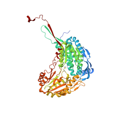
4LEM

Crystal structure of the Delta-pyrroline-5-carboxylate dehydrogenase from Mycobacterium tuberculosis


Experimental Data Snapshot

  • Method: X-RAY DIFFRACTION
  • Resolution: 2.27 Å
  • R-Value Free: 0.204 
  • R-Value Work: 0.148 
  • R-Value Observed: 0.151 

wwPDB Validation   3D Report Full Report


Ligand Structure Quality Assessment 


This is version 1.3 of the entry. See complete history


Literature

Use of a "silver bullet" to resolve crystal lattice dislocation disorder: A cobalamin complex of Delta (1)-pyrroline-5-carboxylate dehydrogenase from Mycobacterium tuberculosis.

Lagautriere, T.Bashiri, G.Baker, E.N.

(2015) J Struct Biol 189: 153-157

  • DOI: https://doi.org/10.1016/j.jsb.2014.12.007
  • Primary Citation of Related Structures:  
    4LEM, 4NS3

  • PubMed Abstract: 

    The use of small molecules as "silver bullets" that can bind to generate crosslinks between protein molecules has been advanced as a powerful means of enhancing success in protein crystallization (McPherson and Cudney, 2006). We have explored this approach in attempts to overcome an order-disorder phenomenon that complicated the structural analysis of the enzyme Δ(1)-pyrroline-5-carboxylate dehydrogenase from Mycobacterium tuberculosis (P5CDH, Mtb-PruA). Using the Silver Bullets Bio screen, we obtained new crystal packing using cobalamin as a co-crystallization agent. This crystal form did not display the order-disorder phenomenon previously encountered. Solution of the crystal structure showed that cobalamin molecules are present in the crystal contacts. Although the cobalamin binding probably does not have physiological relevance, it reflects similarities in the nucleotide-binding region of Mtb-PruA, with the nucleotide loop of cobalamin sharing the binding site for the adenine moiety of NAD(+).


  • Organizational Affiliation

    Structural Biology Laboratory, School of Biological Sciences and Maurice Wilkins Centre for Molecular Biodiscovery, The University of Auckland, Auckland 1010, New Zealand.


Macromolecules
Find similar proteins by:  (by identity cutoff)  |  3D Structure
Entity ID: 1
MoleculeChains Sequence LengthOrganismDetailsImage
1-pyrroline-5-carboxylate dehydrogenase
A, B, C, D, E
A, B, C, D, E, F
563Mycobacterium tuberculosis H37RvMutation(s): 1 
Gene Names: RVBD_1187
EC: 1.5.1.12
UniProt
Find proteins for O50443 (Mycobacterium tuberculosis (strain ATCC 25618 / H37Rv))
Explore O50443 
Go to UniProtKB:  O50443
Entity Groups  
Sequence Clusters30% Identity50% Identity70% Identity90% Identity95% Identity100% Identity
UniProt GroupO50443
Sequence Annotations
Expand
  • Reference Sequence
Experimental Data & Validation

Experimental Data

  • Method: X-RAY DIFFRACTION
  • Resolution: 2.27 Å
  • R-Value Free: 0.204 
  • R-Value Work: 0.148 
  • R-Value Observed: 0.151 
  • Space Group: P 31 2 1
Unit Cell:
Length ( Å )Angle ( ˚ )
a = 164.299α = 90
b = 164.299β = 90
c = 259.114γ = 120
Software Package:
Software NamePurpose
Blu-Icedata collection
PHASERphasing
REFMACrefinement
XDSdata reduction
SCALAdata scaling

Structure Validation

View Full Validation Report


Ligand Structure Quality Assessment 


Entry History 

Deposition Data

Revision History  (Full details and data files)

  • Version 1.0: 2014-05-14
    Type: Initial release
  • Version 1.1: 2014-05-21
    Changes: Database references
  • Version 1.2: 2014-11-12
    Changes: Structure summary
  • Version 1.3: 2015-03-04
    Changes: Database references