Use of a "silver bullet" to resolve crystal lattice dislocation disorder: A cobalamin complex of Delta (1)-pyrroline-5-carboxylate dehydrogenase from Mycobacterium tuberculosis.
Lagautriere, T., Bashiri, G., Baker, E.N.(2015) J Struct Biol 189: 153-157
- PubMed: 25557497 Search on PubMed
- DOI: https://doi.org/10.1016/j.jsb.2014.12.007
- Primary Citation Related Structures:
4LEM, 4NS3 - PubMed Abstract:
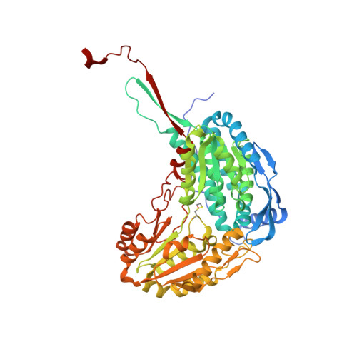
The use of small molecules as "silver bullets" that can bind to generate crosslinks between protein molecules has been advanced as a powerful means of enhancing success in protein crystallization (McPherson and Cudney, 2006). We have explored this approach in attempts to overcome an order-disorder phenomenon that complicated the structural analysis of the enzyme Δ(1)-pyrroline-5-carboxylate dehydrogenase from Mycobacterium tuberculosis (P5CDH, Mtb-PruA). Using the Silver Bullets Bio screen, we obtained new crystal packing using cobalamin as a co-crystallization agent. This crystal form did not display the order-disorder phenomenon previously encountered. Solution of the crystal structure showed that cobalamin molecules are present in the crystal contacts. Although the cobalamin binding probably does not have physiological relevance, it reflects similarities in the nucleotide-binding region of Mtb-PruA, with the nucleotide loop of cobalamin sharing the binding site for the adenine moiety of NAD(+).
- Structural Biology Laboratory, School of Biological Sciences and Maurice Wilkins Centre for Molecular Biodiscovery, The University of Auckland, Auckland 1010, New Zealand.
Organizational Affiliation: