4L6G

Crystal Structure of P450cin Y81F mutant, crystallized in 7 mM 1,8-cineole


Experimental Data Snapshot

  • Method: X-RAY DIFFRACTION
  • Resolution: 1.37 Å
  • R-Value Free: 0.207 
  • R-Value Work: 0.167 
  • R-Value Observed: 0.169 

wwPDB Validation   3D Report Full Report


Ligand Structure Quality Assessment 


This is version 1.2 of the entry. See complete history


Literature

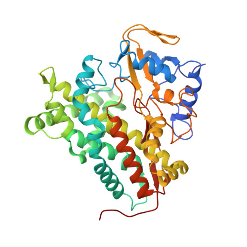
P450cin active site water: implications for substrate binding and solvent accessibility.

Madrona, Y.Hollingsworth, S.A.Khan, B.Poulos, T.L.

(2013) Biochemistry 52: 5039-5050

  • DOI: https://doi.org/10.1021/bi4006946
  • Primary Citation of Related Structures:  
    4L6G, 4L77, 4LHT

  • PubMed Abstract: 
  • In P450cin, Tyr81, Asp241, Asn242, two water molecules, and the substrate participate in a complex H-bonded network. The role of this H-bonded network in substrate binding and catalysis has been probed by crystallography, spectroscopy, kinetics, isothermal titration calorimetry (ITC), and molecular dynamics ...

    In P450cin, Tyr81, Asp241, Asn242, two water molecules, and the substrate participate in a complex H-bonded network. The role of this H-bonded network in substrate binding and catalysis has been probed by crystallography, spectroscopy, kinetics, isothermal titration calorimetry (ITC), and molecular dynamics. For the Y81F mutant, the substrate binds about 20-fold more weakly and Vmax decreases by about 30% in comparison to WT. The enhanced susceptibility of the heme to H₂O₂-mediated destruction in Y81F suggests that this mutant favors the open, low-spin conformational state. Asn242 H-bonds directly with the substrate, and replacing this residue with Ala results in water taking the place of the missing Asn side chain. This mutant exhibits a 70% decrease in activity. Crystal structures and molecular dynamics simulations of substrate-bound complexes show that the solvent has more ready access to the active site, especially for the N242A mutant. This accounts for about a 64% uncoupling of electron transfer from substrate hydroxylation. These data indicate the importance of the interconnected water network on substrate binding and on the open/closed conformational equilibrium, which are both critically important for maintaining high-coupling efficiency.


    Organizational Affiliation

    Department of Molecular Biology and Biochemistry, University of California, Irvine, California 92697-3900, United States.


Macromolecules
Find similar proteins by:  (by identity cutoff)  |  3D Structure
Entity ID: 1
MoleculeChainsSequence LengthOrganismDetailsImage
P450cin
A, B
398Citrobacter braakiiMutation(s): 1 
Gene Names: CIN AcinA
EC: 1.14 (PDB Primary Data), 1.14.14.133 (UniProt)
UniProt
Find proteins for Q8VQF6 (Citrobacter braakii)
Explore Q8VQF6 
Go to UniProtKB:  Q8VQF6
Entity Groups  
Sequence Clusters30% Identity50% Identity70% Identity90% Identity95% Identity100% Identity
UniProt GroupQ8VQF6
Protein Feature View
Expand
  • Reference Sequence
Small Molecules
Ligands 4 Unique
IDChainsName / Formula / InChI Key2D Diagram3D Interactions
HEM
Query on HEM

Download Ideal Coordinates CCD File 
C [auth A],
G [auth B]
PROTOPORPHYRIN IX CONTAINING FE
C34 H32 Fe N4 O4
KABFMIBPWCXCRK-RGGAHWMASA-L
 Ligand Interaction
CNL
Query on CNL

Download Ideal Coordinates CCD File 
D [auth A],
H [auth B]
1,3,3-TRIMETHYL-2-OXABICYCLO[2.2.2]OCTANE
C10 H18 O
WEEGYLXZBRQIMU-WAAGHKOSSA-N
 Ligand Interaction
PGE
Query on PGE

Download Ideal Coordinates CCD File 
F [auth A],
K [auth B]
TRIETHYLENE GLYCOL
C6 H14 O4
ZIBGPFATKBEMQZ-UHFFFAOYSA-N
 Ligand Interaction
SO4
Query on SO4

Download Ideal Coordinates CCD File 
E [auth A],
I [auth B],
J [auth B]
SULFATE ION
O4 S
QAOWNCQODCNURD-UHFFFAOYSA-L
 Ligand Interaction
Binding Affinity Annotations 
IDSourceBinding Affinity
CNL Binding MOAD:  4L6G Kd: 1.61e+5 (nM) from 1 assay(s)
Experimental Data & Validation

Experimental Data

  • Method: X-RAY DIFFRACTION
  • Resolution: 1.37 Å
  • R-Value Free: 0.207 
  • R-Value Work: 0.167 
  • R-Value Observed: 0.169 
  • Space Group: P 21 21 21
Unit Cell:
Length ( Å )Angle ( ˚ )
a = 68.81α = 90
b = 103.77β = 90
c = 127.71γ = 90
Software Package:
Software NamePurpose
MOSFLMdata reduction
PHASERphasing
PHENIXrefinement
SCALAdata scaling

Structure Validation

View Full Validation Report


Ligand Structure Quality Assessment 


Entry History 

Deposition Data

Revision History  (Full details and data files)

  • Version 1.0: 2013-07-24
    Type: Initial release
  • Version 1.1: 2013-08-14
    Changes: Database references
  • Version 1.2: 2023-09-20
    Changes: Data collection, Database references, Derived calculations, Refinement description