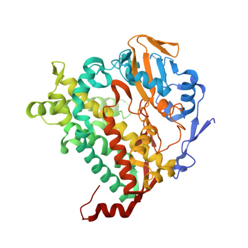
4L0F

Structure of P450sky (CYP163B3), a cytochrome P450 from skyllamycin biosynthesis (open active site)


Experimental Data Snapshot

  • Method: X-RAY DIFFRACTION
  • Resolution: 1.90 Å
  • R-Value Free: 0.221 
  • R-Value Work: 0.179 
  • R-Value Observed: 0.181 

Starting Model: experimental
View more details

wwPDB Validation   3D Report Full Report


Ligand Structure Quality Assessment 


This is version 1.3 of the entry. See complete history


Literature

Cytochrome p450sky interacts directly with the nonribosomal Peptide synthetase to generate three amino Acid precursors in skyllamycin biosynthesis.

Uhlmann, S.Sussmuth, R.D.Cryle, M.J.

(2013) ACS Chem Biol 8: 2586-2596

  • DOI: https://doi.org/10.1021/cb400555e
  • Primary Citation of Related Structures:  
    4L0E, 4L0F

  • PubMed Abstract: 

    The generation of modified amino acid precursors for incorporation in nonribosomal peptide synthesis (NRPS) plays a crucial, if often understated, role in the generation of peptide natural products. The biosynthesis of the cyclic depsipeptide skyllamycin requires three β-hydroxylated amino acid precursors, with in vivo gene inactivation experiments implicating cytochrome P450sky (CYP163B3) in the hydroxylation of these amino acids. Here, we demonstrate the in vitro oxidation of l-amino acid substrates bound to peptidyl carrier protein (PCP) domains 5, 7, and 11 of the skyllamycin nonribosomal synthetase by P450sky. Selectivity for these domains over other PCP domains could be demonstrated, with hydroxylation selective for l-amino acids and stereospecific in nature resulting in the (2S,3S)-configuration. The oxidation of amino acids or small molecule substrate analogues was not supported, demonstrating the necessity of the carrier protein in P450sky-catalyzed hydroxylation. The binding of aminoacyl-PCP substrates to P450sky was detected for the catalytically active PCP7 but not for the catalytically inactive PCP10, indicating carrier protein-mediated selectivity in P450sky substrate binding. X-ray crystal structures of P450sky reveal a 3D-structure with a highly open active site, the size of which is dictated by the carrier protein bound nature of the substrate. P450sky is the first P450 demonstrated to not only interact directly with PCP-bound amino acids within the peptide-forming NRPS but also to do so with three different PCP domains in a specific fashion. This represents an expansion of the complexity and scope of NRPS-mediated peptide synthesis, with the generation of hydroxylated amino acid precursors occurring through the interaction of P450 enzymes following, rather than prior to, the selection of amino acids by NRPS-adenylation domains.


  • Organizational Affiliation

    Institut für Chemie, Technische Universität Berlin , Strasse des 17. Juni 124, 10623 Berlin, Germany.


Macromolecules
Find similar proteins by:  (by identity cutoff)  |  3D Structure
Entity ID: 1
MoleculeChains Sequence LengthOrganismDetailsImage
P450 monooxygenase446Streptomyces sp. Acta 2897Mutation(s): 0 
Gene Names: sky32
EC: 1.14
UniProt
Find proteins for F2YRY7 (Streptomyces sp. Acta 2897)
Explore F2YRY7 
Go to UniProtKB:  F2YRY7
Entity Groups  
Sequence Clusters30% Identity50% Identity70% Identity90% Identity95% Identity100% Identity
UniProt GroupF2YRY7
Sequence Annotations
Expand
  • Reference Sequence
Small Molecules
Ligands 3 Unique
IDChains Name / Formula / InChI Key2D Diagram3D Interactions
HEM
Query on HEM

Download Ideal Coordinates CCD File 
B [auth A]PROTOPORPHYRIN IX CONTAINING FE
C34 H32 Fe N4 O4
KABFMIBPWCXCRK-RGGAHWMASA-L
PGE
Query on PGE

Download Ideal Coordinates CCD File 
Q [auth A]TRIETHYLENE GLYCOL
C6 H14 O4
ZIBGPFATKBEMQZ-UHFFFAOYSA-N
EDO
Query on EDO

Download Ideal Coordinates CCD File 
C [auth A]
D [auth A]
E [auth A]
F [auth A]
G [auth A]
C [auth A],
D [auth A],
E [auth A],
F [auth A],
G [auth A],
H [auth A],
I [auth A],
J [auth A],
K [auth A],
L [auth A],
M [auth A],
N [auth A],
O [auth A],
P [auth A]
1,2-ETHANEDIOL
C2 H6 O2
LYCAIKOWRPUZTN-UHFFFAOYSA-N
Experimental Data & Validation

Experimental Data

  • Method: X-RAY DIFFRACTION
  • Resolution: 1.90 Å
  • R-Value Free: 0.221 
  • R-Value Work: 0.179 
  • R-Value Observed: 0.181 
  • Space Group: P 1 21 1
Unit Cell:
Length ( Å )Angle ( ˚ )
a = 48.02α = 90
b = 79.55β = 104.53
c = 62.88γ = 90
Software Package:
Software NamePurpose
XSCALEdata scaling
PHASERphasing
REFMACrefinement
PDB_EXTRACTdata extraction
XDSdata scaling

Structure Validation

View Full Validation Report


Ligand Structure Quality Assessment 


Entry History 

Deposition Data

  • Released Date: 2013-09-25 
  • Deposition Author(s): Cryle, M.J.

Revision History  (Full details and data files)

  • Version 1.0: 2013-09-25
    Type: Initial release
  • Version 1.1: 2013-10-16
    Changes: Database references
  • Version 1.2: 2013-11-27
    Changes: Database references
  • Version 1.3: 2023-09-20
    Changes: Data collection, Database references, Derived calculations, Refinement description