4K2G

Structure of Pseudomonas aeruginosa PvdQ bound to BRD-A33442372


Experimental Data Snapshot

  • Method: X-RAY DIFFRACTION
  • Resolution: 2.30 Å
  • R-Value Free: 0.248 
  • R-Value Work: 0.191 
  • R-Value Observed: 0.194 

wwPDB Validation   3D Report Full Report


Ligand Structure Quality Assessment 


This is version 1.2 of the entry. See complete history


Literature

Identification of Inhibitors of PvdQ, an Enzyme Involved in the Synthesis of the Siderophore Pyoverdine.

Wurst, J.M.Drake, E.J.Theriault, J.R.Jewett, I.T.VerPlank, L.Perez, J.R.Dandapani, S.Palmer, M.Moskowitz, S.M.Schreiber, S.L.Munoz, B.Gulick, A.M.

(2014) ACS Chem Biol 9: 1536-1544

  • DOI: https://doi.org/10.1021/cb5001586
  • Primary Citation of Related Structures:  
    4K2F, 4K2G

  • PubMed Abstract: 

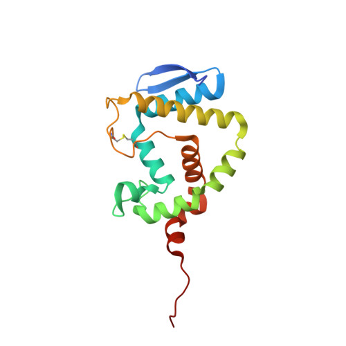
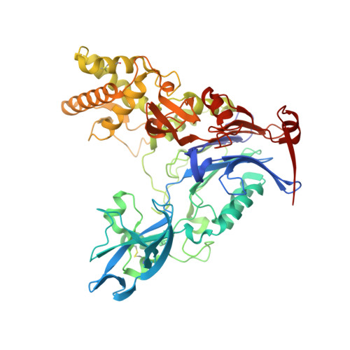
    Pseudomonas aeruginosa produces the peptide siderophore pyoverdine, which is used to acquire essential Fe(3+) ions from the environment. PvdQ, an Ntn hydrolase, is required for the biosynthesis of pyoverdine. PvdQ knockout strains are not infectious in model systems, suggesting that disruption of siderophore production via PvdQ inhibition could be exploited as a target for novel antibacterial agents, by preventing cells from acquiring iron in the low iron environments of most biological settings. We have previously described a high-throughput screen to identify inhibitors of PvdQ that identified inhibitors with IC50 values of ∼100 μM. Here, we describe the discovery of ML318, a biaryl nitrile inhibitor of PvdQ acylase. ML318 inhibits PvdQ in vitro (IC50 = 20 nM) by binding in the acyl-binding site, as confirmed by the X-ray crystal structure of PvdQ bound to ML318. Additionally, the PvdQ inhibitor is active in a whole cell assay, preventing pyoverdine production and limiting the growth of P. aeruginosa under iron-limiting conditions.


  • Organizational Affiliation

    The Broad Institute , Cambridge, Massachusetts 02142, United States.


Macromolecules
Find similar proteins by:  (by identity cutoff)  |  3D Structure
Entity ID: 1
MoleculeChains Sequence LengthOrganismDetailsImage
Acyl-homoserine lactone acylase PvdQ170Pseudomonas aeruginosa PAO1Mutation(s): 0 
Gene Names: pvdQqsc112PA2385
EC: 3.5.1.97
UniProt
Find proteins for Q9I194 (Pseudomonas aeruginosa (strain ATCC 15692 / DSM 22644 / CIP 104116 / JCM 14847 / LMG 12228 / 1C / PRS 101 / PAO1))
Explore Q9I194 
Go to UniProtKB:  Q9I194
Entity Groups  
Sequence Clusters30% Identity50% Identity70% Identity90% Identity95% Identity100% Identity
UniProt GroupQ9I194
Sequence Annotations
Expand
  • Reference Sequence
Find similar proteins by:  (by identity cutoff)  |  3D Structure
Entity ID: 2
MoleculeChains Sequence LengthOrganismDetailsImage
Acyl-homoserine lactone acylase PvdQ546Pseudomonas aeruginosa PAO1Mutation(s): 0 
Gene Names: pvdQqsc112PA2385
EC: 3.5.1.97
UniProt
Find proteins for Q9I194 (Pseudomonas aeruginosa (strain ATCC 15692 / DSM 22644 / CIP 104116 / JCM 14847 / LMG 12228 / 1C / PRS 101 / PAO1))
Explore Q9I194 
Go to UniProtKB:  Q9I194
Entity Groups  
Sequence Clusters30% Identity50% Identity70% Identity90% Identity95% Identity100% Identity
UniProt GroupQ9I194
Sequence Annotations
Expand
  • Reference Sequence
Small Molecules
Ligands 2 Unique
IDChains Name / Formula / InChI Key2D Diagram3D Interactions
1OQ
Query on 1OQ

Download Ideal Coordinates CCD File 
Q [auth B](2S)-(4-fluorophenyl)[6-(trifluoromethyl)pyridin-2-yl]ethanenitrile
C14 H8 F4 N2
DXQNDKQUHKVTTC-NSHDSACASA-N
EDO
Query on EDO

Download Ideal Coordinates CCD File 
C [auth A]
D [auth A]
E [auth A]
F [auth A]
G [auth A]
C [auth A],
D [auth A],
E [auth A],
F [auth A],
G [auth A],
H [auth B],
I [auth B],
J [auth B],
K [auth B],
L [auth B],
M [auth B],
N [auth B],
O [auth B],
P [auth B]
1,2-ETHANEDIOL
C2 H6 O2
LYCAIKOWRPUZTN-UHFFFAOYSA-N
Binding Affinity Annotations 
IDSourceBinding Affinity
1OQ Binding MOAD:  4K2G IC50: 20 (nM) from 1 assay(s)
BindingDB:  4K2G IC50: 20 (nM) from 1 assay(s)
Experimental Data & Validation

Experimental Data

  • Method: X-RAY DIFFRACTION
  • Resolution: 2.30 Å
  • R-Value Free: 0.248 
  • R-Value Work: 0.191 
  • R-Value Observed: 0.194 
  • Space Group: C 2 2 21
Unit Cell:
Length ( Å )Angle ( ˚ )
a = 120.335α = 90
b = 165.508β = 90
c = 93.988γ = 90
Software Package:
Software NamePurpose
CrystalCleardata collection
MOLREPphasing
REFMACrefinement
HKL-2000data reduction
HKL-2000data scaling

Structure Validation

View Full Validation Report


Ligand Structure Quality Assessment 


Entry History 

Deposition Data

Revision History  (Full details and data files)

  • Version 1.0: 2014-06-18
    Type: Initial release
  • Version 1.1: 2014-07-30
    Changes: Database references
  • Version 1.2: 2023-09-20
    Changes: Data collection, Database references, Derived calculations, Refinement description