4JLW

Crystal structure of formaldehyde dehydrogenase from Pseudomonas aeruginosa


Experimental Data Snapshot

  • Method: X-RAY DIFFRACTION
  • Resolution: 2.70 Å
  • R-Value Free: 0.259 
  • R-Value Work: 0.209 
  • R-Value Observed: 0.212 

wwPDB Validation   3D Report Full Report


Ligand Structure Quality Assessment 


This is version 1.1 of the entry. See complete history


Literature

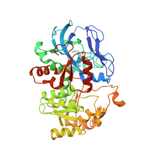
Structure of formaldehyde dehydrogenase from Pseudomonas aeruginosa: the binary complex with the cofactor NAD+.

Liao, Y.P.Chen, S.Wang, D.L.Zhang, W.Wang, S.Ding, J.F.Wang, Y.M.Cai, L.J.Ran, X.Y.Wang, X.Zhu, H.X.

(2013) Acta Crystallogr Sect F Struct Biol Cryst Commun 69: 967-972

  • DOI: https://doi.org/10.1107/S174430911302160X
  • Primary Citation of Related Structures:  
    4JLW

  • PubMed Abstract: 

    Formaldehyde dehydrogenase (FDH) is a member of the zinc-containing medium-chain alcohol dehydrogenase family which oxidizes toxic formaldehyde to formate using NAD(+) as an electron carrier. Three-dimensional structures have been reported for FDHs from several different species. Most FDHs are dependent on glutathione for catalysis, but the enzyme from Pseudomonas putida is an exception. In this structural communication, the recombinant production, crystallization and X-ray structure determination at 2.7 Å resolution of FDH from P. aeruginosa are described. Both the tetrameric assembly and the NAD(+)-binding mode of P. aeruginosa FDH are similar to those of P. putida FDH, which is in good agreement with the high sequence identity (87.97%) between these two proteins. Preliminary enzymatic kinetics studies of P. aeruginosa FDH also revealed a conserved glutathione-independent `ping-pong' mechanism of formaldehyde oxidization.


  • Organizational Affiliation

    R&D Department, Novoprotein Scientific Inc. (Shanghai), R202, Building 2, 720 Cailun Road, Shanghai 201203, People's Republic of China.


Macromolecules
Find similar proteins by:  (by identity cutoff)  |  3D Structure
Entity ID: 1
MoleculeChains Sequence LengthOrganismDetailsImage
Glutathione-independent formaldehyde dehydrogenase
A, B, C, D
399Pseudomonas aeruginosa LESB58Mutation(s): 0 
Gene Names: fdhAPLES_58161
Entity Groups  
Sequence Clusters30% Identity50% Identity70% Identity90% Identity95% Identity100% Identity
Sequence Annotations
Expand
  • Reference Sequence
Small Molecules
Ligands 3 Unique
IDChains Name / Formula / InChI Key2D Diagram3D Interactions
NAD
Query on NAD

Download Ideal Coordinates CCD File 
G [auth A],
K [auth B],
P [auth C],
U [auth D]
NICOTINAMIDE-ADENINE-DINUCLEOTIDE
C21 H27 N7 O14 P2
BAWFJGJZGIEFAR-NNYOXOHSSA-N
SO4
Query on SO4

Download Ideal Coordinates CCD File 
H [auth A]
L [auth B]
M [auth B]
Q [auth C]
R [auth C]
H [auth A],
L [auth B],
M [auth B],
Q [auth C],
R [auth C],
V [auth D]
SULFATE ION
O4 S
QAOWNCQODCNURD-UHFFFAOYSA-L
ZN
Query on ZN

Download Ideal Coordinates CCD File 
E [auth A]
F [auth A]
I [auth B]
J [auth B]
N [auth C]
E [auth A],
F [auth A],
I [auth B],
J [auth B],
N [auth C],
O [auth C],
S [auth D],
T [auth D]
ZINC ION
Zn
PTFCDOFLOPIGGS-UHFFFAOYSA-N
Experimental Data & Validation

Experimental Data

  • Method: X-RAY DIFFRACTION
  • Resolution: 2.70 Å
  • R-Value Free: 0.259 
  • R-Value Work: 0.209 
  • R-Value Observed: 0.212 
  • Space Group: P 1 21 1
Unit Cell:
Length ( Å )Angle ( ˚ )
a = 89.226α = 90
b = 98.305β = 91.29
c = 99.623γ = 90
Software Package:
Software NamePurpose
HKL-2000data collection
PHASERphasing
REFMACrefinement
DENZOdata reduction
SCALEPACKdata scaling

Structure Validation

View Full Validation Report


Ligand Structure Quality Assessment 


Entry History 

Deposition Data

Revision History  (Full details and data files)

  • Version 1.0: 2013-10-30
    Type: Initial release
  • Version 1.1: 2023-11-08
    Changes: Data collection, Database references, Derived calculations, Refinement description