Identification of the First PPAR alpha / gamma Dual Agonist Able To Bind to Canonical and Alternative Sites of PPAR gamma and To Inhibit Its Cdk5-Mediated Phosphorylation.
Laghezza, A., Piemontese, L., Cerchia, C., Montanari, R., Capelli, D., Giudici, M., Crestani, M., Tortorella, P., Peiretti, F., Pochetti, G., Lavecchia, A., Loiodice, F.(2018) J Med Chem 61: 8282-8298
- PubMed: 30199253 Search on PubMed
- DOI: https://doi.org/10.1021/acs.jmedchem.8b00835
- Primary Citation Related Structures:
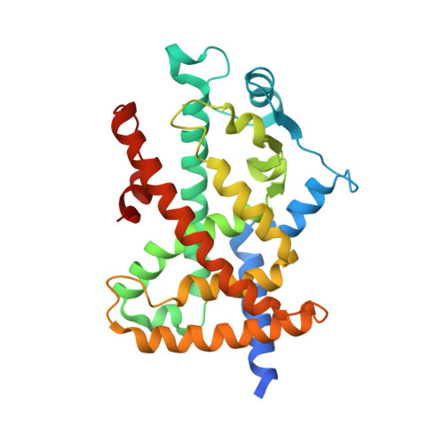
4JL4, 6F2L - PubMed Abstract:
A new series of derivatives of the PPARα/γ dual agonist 1 allowed us to identify the ligand ( S)-6 as a potent partial agonist of both PPARα and γ subtypes. X-ray studies in PPARγ revealed two different binding modes of ( S)-6 to the canonical site. However, ( S)-6 was also able to bind an alternative site as demonstrated by transactivation assay in the presence of a canonical PPARγ antagonist and supported from docking experiments. This compound did not activate the PPARγ-dependent program of adipocyte differentiation inducing a very less severe lipid accumulation compared to rosiglitazone but increased the insulin-stimulated glucose uptake in 3T3-L1 adipocytes. Finally, ( S)-6 inhibited the Cdk5-mediated phosphorylation of PPARγ at serine 273 that is currently considered the mechanism by which some PPARγ partial agonists exert antidiabetic effects similar to thiazolidinediones, without showing their typical side effects. This is the first PPARα/γ dual agonist reported to show this inhibitory effect representing the potential lead of a new class of drugs for treatment of dyslipidemic type 2 diabetes.
- Dipartimento Farmacia-Scienze del Farmaco , Università degli Studi di Bari "Aldo Moro" , Via Orabona 4 , 70125 Bari , Italy.
Organizational Affiliation: