4JKT

Crystal structure of mouse Glutaminase C, BPTES-bound form


Experimental Data Snapshot

  • Method: X-RAY DIFFRACTION
  • Resolution: 2.77 Å
  • R-Value Free: 0.295 
  • R-Value Work: 0.253 
  • R-Value Observed: 0.255 

wwPDB Validation   3D Report Full Report


Ligand Structure Quality Assessment 


This is version 1.4 of the entry. See complete history


Literature

Active Glutaminase C Self-assembles into a Supratetrameric Oligomer That Can Be Disrupted by an Allosteric Inhibitor.

Ferreira, A.P.Cassago, A.Goncalves Kde, A.Dias, M.M.Adamoski, D.Ascencao, C.F.Honorato, R.V.de Oliveira, J.F.Ferreira, I.M.Fornezari, C.Bettini, J.Oliveira, P.S.Paes Leme, A.F.Portugal, R.V.Ambrosio, A.L.Dias, S.M.

(2013) J Biol Chem 288: 28009-28020

  • DOI: https://doi.org/10.1074/jbc.M113.501346
  • Primary Citation of Related Structures:  
    4JKT

  • PubMed Abstract: 

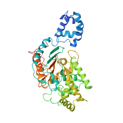
    The phosphate-dependent transition between enzymatically inert dimers into catalytically capable tetramers has long been the accepted mechanism for the glutaminase activation. Here, we demonstrate that activated glutaminase C (GAC) self-assembles into a helical, fiber-like double-stranded oligomer and propose a molecular model consisting of seven tetramer copies per turn per strand interacting via the N-terminal domains. The loop (321)LRFNKL(326) is projected as the major regulating element for self-assembly and enzyme activation. Furthermore, the previously identified in vivo lysine acetylation (Lys(311) in humans, Lys(316) in mouse) is here proposed as an important down-regulator of superoligomer assembly and protein activation. Bis-2-(5-phenylacetamido-1,3,4-thiadiazol-2-yl)ethyl sulfide, a known glutaminase inhibitor, completely disrupted the higher order oligomer, explaining its allosteric mechanism of inhibition via tetramer stabilization. A direct correlation between the tendency to self-assemble and the activity levels of the three mammalian glutaminase isozymes was established, with GAC being the most active enzyme while forming the longest structures. Lastly, the ectopic expression of a fiber-prone superactive GAC mutant in MDA-MB 231 cancer cells provided considerable proliferative advantages to transformed cells. These findings yield unique implications for the development of GAC-oriented therapeutics targeting tumor metabolism.


  • Organizational Affiliation

    From the Laboratórios Nacionais de Biociências e.


Macromolecules
Find similar proteins by:  (by identity cutoff)  |  3D Structure
Entity ID: 1
MoleculeChains Sequence LengthOrganismDetailsImage
Glutaminase kidney isoform, mitochondrial
A, B, C, D
479Mus musculusMutation(s): 0 
Gene Names: GlsGls1Kiaa0838
EC: 3.5.1.2
UniProt & NIH Common Fund Data Resources
Find proteins for D3Z7P3 (Mus musculus)
Explore D3Z7P3 
Go to UniProtKB:  D3Z7P3
IMPC:  MGI:95752
Entity Groups  
Sequence Clusters30% Identity50% Identity70% Identity90% Identity95% Identity100% Identity
UniProt GroupD3Z7P3
Sequence Annotations
Expand
  • Reference Sequence
Small Molecules
Ligands 1 Unique
IDChains Name / Formula / InChI Key2D Diagram3D Interactions
04A
Query on 04A

Download Ideal Coordinates CCD File 
E [auth B],
F [auth D]
N,N'-[sulfanediylbis(ethane-2,1-diyl-1,3,4-thiadiazole-5,2-diyl)]bis(2-phenylacetamide)
C24 H24 N6 O2 S3
MDJIPXYRSZHCFS-UHFFFAOYSA-N
Binding Affinity Annotations 
IDSourceBinding Affinity
04A BindingDB:  4JKT Kd: min: 200, max: 4300 (nM) from 2 assay(s)
IC50: min: 25, max: 8600 (nM) from 12 assay(s)
PDBBind:  4JKT IC50: 80.4 (nM) from 1 assay(s)
Experimental Data & Validation

Experimental Data

  • Method: X-RAY DIFFRACTION
  • Resolution: 2.77 Å
  • R-Value Free: 0.295 
  • R-Value Work: 0.253 
  • R-Value Observed: 0.255 
  • Space Group: P 21 21 21
Unit Cell:
Length ( Å )Angle ( ˚ )
a = 100.67α = 90
b = 140.24β = 90
c = 180.89γ = 90
Software Package:
Software NamePurpose
MAR345data collection
PHASERphasing
PHENIXrefinement
MOSFLMdata reduction
Aimlessdata scaling

Structure Validation

View Full Validation Report


Ligand Structure Quality Assessment 


Entry History 

Deposition Data

Revision History  (Full details and data files)

  • Version 1.0: 2013-08-14
    Type: Initial release
  • Version 1.1: 2013-08-28
    Changes: Database references
  • Version 1.2: 2013-10-16
    Changes: Database references
  • Version 1.3: 2017-11-15
    Changes: Advisory, Refinement description
  • Version 1.4: 2023-09-20
    Changes: Advisory, Data collection, Database references, Derived calculations, Refinement description