4JC5

Immune activator bound to receptor


Experimental Data Snapshot

  • Method: X-RAY DIFFRACTION
  • Resolution: 2.75 Å
  • R-Value Free: 0.237 
  • R-Value Work: 0.209 
  • R-Value Observed: 0.211 

wwPDB Validation   3D Report Full Report


Ligand Structure Quality Assessment 


This is version 1.3 of the entry. See complete history


Literature

Species-specific detection of the antiviral small-molecule compound CMA by STING.

Cavlar, T.Deimling, T.Ablasser, A.Hopfner, K.P.Hornung, V.

(2013) EMBO J 32: 1440-1450

  • DOI: https://doi.org/10.1038/emboj.2013.86
  • Primary Citation of Related Structures:  
    4JC5

  • PubMed Abstract: 

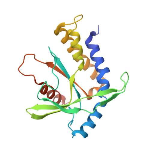
    Extensive research on antiviral small molecules starting in the early 1970s has led to the identification of 10-carboxymethyl-9-acridanone (CMA) as a potent type I interferon (IFN) inducer. Up to date, the mode of action of this antiviral molecule has remained elusive. Here we demonstrate that CMA mediates a cell-intrinsic type I IFN response, depending on the ER-resident protein STING. CMA directly binds to STING and triggers a strong antiviral response through the TBK1/IRF3 route. Interestingly, while CMA displays extraordinary activity in phosphorylating IRF3 in the murine system, CMA fails to activate human cells that are otherwise responsive to STING ligands. This failure to activate human STING can be ascribed to its inability to bind to the C-terminal ligand-binding domain of human STING. Crystallographic studies show that two CMA molecules bind to the central Cyclic diguanylate (c-diGMP)-binding pocket of the STING dimer and fold the lid region in a fashion similar, but partially distinct, to c-diGMP. Altogether, these results provide novel insight into ligand-sensing properties of STING and, furthermore, unravel unexpected species-specific differences of this innate sensor.


  • Organizational Affiliation

    Unit for Clinical Biochemistry, Institute for Clinical Chemistry and Clinical Pharmacology, University Hospital, University of Bonn, Bonn, Germany.


Macromolecules
Find similar proteins by:  (by identity cutoff)  |  3D Structure
Entity ID: 1
MoleculeChains Sequence LengthOrganismDetailsImage
Transmembrane protein 173
A, B
200Mus musculusMutation(s): 0 
Gene Names: Tmem173Eris MitaMpysSting
UniProt & NIH Common Fund Data Resources
Find proteins for Q3TBT3 (Mus musculus)
Explore Q3TBT3 
Go to UniProtKB:  Q3TBT3
IMPC:  MGI:1919762
Entity Groups  
Sequence Clusters30% Identity50% Identity70% Identity90% Identity95% Identity100% Identity
UniProt GroupQ3TBT3
Sequence Annotations
Expand
  • Reference Sequence
Small Molecules
Ligands 1 Unique
IDChains Name / Formula / InChI Key2D Diagram3D Interactions
1K5
Query on 1K5

Download Ideal Coordinates CCD File 
C [auth A],
D [auth B]
(9-oxoacridin-10(9H)-yl)acetic acid
C15 H11 N O3
UOMKBIIXHQIERR-UHFFFAOYSA-N
Binding Affinity Annotations 
IDSourceBinding Affinity
1K5 BindingDB:  4JC5 Kd: 3300 (nM) from 1 assay(s)
Experimental Data & Validation

Experimental Data

  • Method: X-RAY DIFFRACTION
  • Resolution: 2.75 Å
  • R-Value Free: 0.237 
  • R-Value Work: 0.209 
  • R-Value Observed: 0.211 
  • Space Group: H 3
Unit Cell:
Length ( Å )Angle ( ˚ )
a = 108.54α = 90
b = 108.54β = 90
c = 100.83γ = 120
Software Package:
Software NamePurpose
PHASERphasing
PHENIXrefinement
XDSdata reduction
XDSdata scaling

Structure Validation

View Full Validation Report


Ligand Structure Quality Assessment 


Entry History 

Deposition Data

Revision History  (Full details and data files)

  • Version 1.0: 2013-05-08
    Type: Initial release
  • Version 1.1: 2013-05-29
    Changes: Database references
  • Version 1.2: 2017-11-15
    Changes: Advisory, Refinement description
  • Version 1.3: 2024-02-28
    Changes: Advisory, Data collection, Database references, Derived calculations