4IHV | pdb_00004ihv

Crystal structure of Fis bound to 27 bp sequence DNA F28 (AAATTTGTTTGAGCGTTGAGCAAATTT)


Experimental Data Snapshot

  • Method: X-RAY DIFFRACTION
  • Resolution: 2.72 Å
  • R-Value Free: 
    0.258 (Depositor), 0.261 (DCC) 
  • R-Value Work: 
    0.217 (Depositor), 0.222 (DCC) 
  • R-Value Observed: 
    0.221 (Depositor) 

Starting Model: experimental
View more details

wwPDB Validation 3D Report Full Report

Validation slider image for 4IHV

This is version 1.3 of the entry. See complete history

Literature

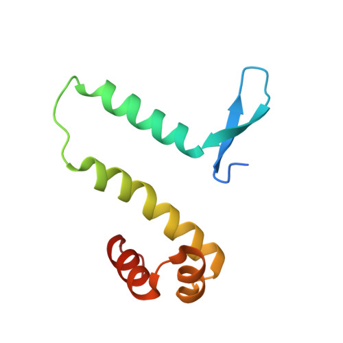
Control of DNA minor groove width and Fis protein binding by the purine 2-amino group.

Hancock, S.P.Ghane, T.Cascio, D.Rohs, R.Di Felice, R.Johnson, R.C.

(2013) Nucleic Acids Res 41: 6750-6760

  • DOI: https://doi.org/10.1093/nar/gkt357
  • Primary Citation Related Structures: 
    4IHV, 4IHW, 4IHX, 4IHY

  • PubMed Abstract: 

    The width of the DNA minor groove varies with sequence and can be a major determinant of DNA shape recognition by proteins. For example, the minor groove within the center of the Fis-DNA complex narrows to about half the mean minor groove width of canonical B-form DNA to fit onto the protein surface. G/C base pairs within this segment, which is not contacted by the Fis protein, reduce binding affinities up to 2000-fold over A/T-rich sequences. We show here through multiple X-ray structures and binding properties of Fis-DNA complexes containing base analogs that the 2-amino group on guanine is the primary molecular determinant controlling minor groove widths. Molecular dynamics simulations of free-DNA targets with canonical and modified bases further demonstrate that sequence-dependent narrowing of minor groove widths is modulated almost entirely by the presence of purine 2-amino groups. We also provide evidence that protein-mediated phosphate neutralization facilitates minor groove compression and is particularly important for binding to non-optimally shaped DNA duplexes.


  • Organizational Affiliation
    • Department of Biological Chemistry, David Geffen School of Medicine at the University of California at Los Angeles, Los Angeles, CA 90095-1737, USA.

Macromolecule Content 

  • Total Structure Weight: 39.09 kDa 
  • Atom Count: 2,611 
  • Modeled Residue Count: 243 
  • Deposited Residue Count: 250 
  • Unique protein chains: 1
  • Unique nucleic acid chains: 2

Macromolecules


Find similar proteins by:|  3D Structure
Entity ID: 1
MoleculeChains  Sequence LengthOrganismDetailsImage
DNA-binding protein fis
A, B
98Escherichia coli K-12Mutation(s): 0 
Gene Names: ECDH1ME8569_3147EcDH1_0445fis
Find similar nucleic acids by:  (by identity cutoff) 
Entity ID: 2
MoleculeChains LengthOrganismImage
27-bp DNA Strand A27N/A
Sequence Annotations
Expand
Reference Sequence
Find similar nucleic acids by:  (by identity cutoff) 
Entity ID: 3
MoleculeChains LengthOrganismImage
27-bp DNA Strand B27N/A
Sequence Annotations
Expand
Reference Sequence

Experimental Data & Validation

Experimental Data

  • Method: X-RAY DIFFRACTION
  • Resolution: 2.72 Å
  • R-Value Free:  0.258 (Depositor), 0.261 (DCC) 
  • R-Value Work:  0.217 (Depositor), 0.222 (DCC) 
  • R-Value Observed: 0.221 (Depositor) 
Space Group: P 21 21 21
Unit Cell:
Length ( Å )Angle ( ˚ )
a = 42.79α = 90
b = 89.39β = 90
c = 154.15γ = 90
Software Package:
Software NamePurpose
XSCALEdata scaling
PHASERphasing
PHENIXrefinement
PDB_EXTRACTdata extraction
XDSdata reduction

Structure Validation

View Full Validation Report


Entry History 

Deposition Data

Revision History  (Full details and data files)

  • Version 1.0: 2013-05-01
    Type: Initial release
  • Version 1.1: 2013-05-22
    Changes: Database references
  • Version 1.2: 2013-07-31
    Changes: Database references
  • Version 1.3: 2023-09-20
    Changes: Data collection, Database references, Refinement description