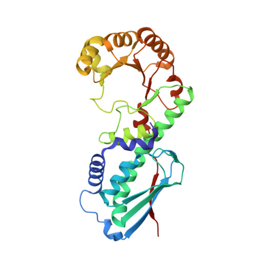
Crystal structure of Sus scrofa quinolinate phosphoribosyltransferase in complex with nicotinate mononucleotide
Youn, H.-S., Kim, M.-K., Kang, G.B., Kim, T.G., Lee, J.-G., An, J.Y., Park, K.R., Lee, Y., Kang, J.Y., Song, H.E., Park, I., Cho, C., Fukuoka, S., Eom, S.H.(2013) PLoS One 8: e62027-e62027
- PubMed: 23626766 Search on PubMedSearch on PubMed Central
- DOI: https://doi.org/10.1371/journal.pone.0062027
- Primary Citation Related Structures:
4I9A - PubMed Abstract:
We have determined the crystal structure of porcine quinolinate phosphoribosyltransferase (QAPRTase) in complex with nicotinate mononucleotide (NAMN), which is the first crystal structure of a mammalian QAPRTase with its reaction product. The structure was determined from protein obtained from the porcine kidney. Because the full protein sequence of porcine QAPRTase was not available in either protein or nucleotide databases, cDNA was synthesized using reverse transcriptase-polymerase chain reaction to determine the porcine QAPRTase amino acid sequence. The crystal structure revealed that porcine QAPRTases have a hexameric structure that is similar to other eukaryotic QAPRTases, such as the human and yeast enzymes. However, the interaction between NAMN and porcine QAPRTase was different from the interaction found in prokaryotic enzymes, such as those of Helicobacter pylori and Mycobacterium tuberculosis. The crystal structure of porcine QAPRTase in complex with NAMN provides a structural framework for understanding the unique properties of the mammalian QAPRTase active site and designing new antibiotics that are selective for the QAPRTases of pathogenic bacteria, such as H. pylori and M. tuberculosis.
- School of Life Sciences, Gwangju Institute of Science & Technology, Gwangju, Republic of Korea.
Organizational Affiliation: