4I1O

Crystal structure of the Legionella pneumophila GAP domain of LepB in complex with Rab1b bound to GDP and BeF3


Experimental Data Snapshot

  • Method: X-RAY DIFFRACTION
  • Resolution: 2.70 Å
  • R-Value Free: 0.249 
  • R-Value Work: 0.217 
  • R-Value Observed: 0.219 

wwPDB Validation   3D Report Full Report


Ligand Structure Quality Assessment 


This is version 1.1 of the entry. See complete history


Literature

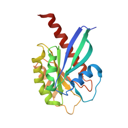
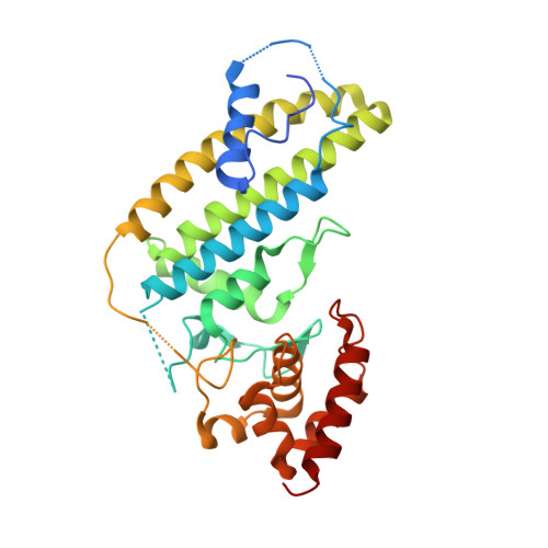
Mechanism of Rab1b deactivation by the Legionella pneumophila GAP LepB.

Mihai Gazdag, E.Streller, A.Haneburger, I.Hilbi, H.Vetter, I.R.Goody, R.S.Itzen, A.

(2013) EMBO Rep 14: 199-205

  • DOI: 10.1038/embor.2012.211
  • Primary Citation of Related Structures:  
    4I1M, 4I1O

  • PubMed Abstract: 
  • Legionella pneumophila is an intracellularly surviving pathogen that releases about 270 different proteins into the host cell during infection. A set of secreted proteins takes control of the vesicular trafficking regulator Rab1. Legionella LepB inactivates Rab1 by acting as a GTPase-activating protein (GAP) ...

    Legionella pneumophila is an intracellularly surviving pathogen that releases about 270 different proteins into the host cell during infection. A set of secreted proteins takes control of the vesicular trafficking regulator Rab1. Legionella LepB inactivates Rab1 by acting as a GTPase-activating protein (GAP). We present the crystal structure of the Rab1b:LepB complex together with a thorough biochemical analysis and show that the GAP domain of LepB consists of an unusual fold. LepB acts by an atypical RabGAP mechanism that is reminiscent of classical GAPs and therefore sets the protein apart from mammalian TBC-like GAPs. Surprisingly, LepB can function as a GAP for Rab3, Rab8, Rab13 and Rab35, too, suggesting that it has a broader cellular role than previously thought.


    Organizational Affiliation

    Max-Planck-Institute of Molecular Physiology, Department of Physical Biochemistry, Dortmund 44227, Germany.


Macromolecules
Find similar proteins by:  (by identity cutoff)  |  3D Structure
Entity ID: 1
MoleculeChainsSequence LengthOrganismDetailsImage
Ras-related protein Rab-1BA, C, E, G181Homo sapiensMutation(s): 0 
Gene Names: RAB1B
EC: 3.6.5.2
UniProt & NIH Common Fund Data Resources
Find proteins for Q9H0U4 (Homo sapiens)
Explore Q9H0U4 
Go to UniProtKB:  Q9H0U4
PHAROS:  Q9H0U4
Entity Groups  
Sequence Clusters30% Identity50% Identity70% Identity90% Identity95% Identity100% Identity
UniProt GroupQ9H0U4
Protein Feature View
Expand
  • Reference Sequence
Find similar proteins by:  (by identity cutoff)  |  3D Structure
Entity ID: 2
MoleculeChainsSequence LengthOrganismDetailsImage
LepBB, D, F, H302Legionella pneumophila subsp. pneumophila str. Philadelphia 1Mutation(s): 3 
Gene Names: lepBlpg2490
UniProt
Find proteins for Q5ZSM7 (Legionella pneumophila subsp. pneumophila (strain Philadelphia 1 / ATCC 33152 / DSM 7513))
Explore Q5ZSM7 
Go to UniProtKB:  Q5ZSM7
Entity Groups  
Sequence Clusters30% Identity50% Identity70% Identity90% Identity95% Identity100% Identity
UniProt GroupQ5ZSM7
Protein Feature View
Expand
  • Reference Sequence
Small Molecules
Ligands 4 Unique
IDChainsName / Formula / InChI Key2D Diagram3D Interactions
GDP
Query on GDP

Download Ideal Coordinates CCD File 
J [auth A],
N [auth C],
U [auth E],
X [auth G]
GUANOSINE-5'-DIPHOSPHATE
C10 H15 N5 O11 P2
QGWNDRXFNXRZMB-UUOKFMHZSA-N
 Ligand Interaction
PEG
Query on PEG

Download Ideal Coordinates CCD File 
L [auth A],
P [auth C],
Q [auth C],
R [auth C],
S [auth D],
L [auth A],
P [auth C],
Q [auth C],
R [auth C],
S [auth D],
Z [auth G]
DI(HYDROXYETHYL)ETHER
C4 H10 O3
MTHSVFCYNBDYFN-UHFFFAOYSA-N
 Ligand Interaction
BEF
Query on BEF

Download Ideal Coordinates CCD File 
K [auth A],
O [auth C],
V [auth E],
Y [auth G]
BERYLLIUM TRIFLUORIDE ION
Be F3
OGIAHMCCNXDTIE-UHFFFAOYSA-K
 Ligand Interaction
MG
Query on MG

Download Ideal Coordinates CCD File 
I [auth A],
M [auth C],
T [auth E],
W [auth G]
MAGNESIUM ION
Mg
JLVVSXFLKOJNIY-UHFFFAOYSA-N
 Ligand Interaction
Experimental Data & Validation

Experimental Data

  • Method: X-RAY DIFFRACTION
  • Resolution: 2.70 Å
  • R-Value Free: 0.249 
  • R-Value Work: 0.217 
  • R-Value Observed: 0.219 
  • Space Group: P 31
Unit Cell:
Length ( Å )Angle ( ˚ )
a = 124.41α = 90
b = 124.41β = 90
c = 147.47γ = 120
Software Package:
Software NamePurpose
XSCALEdata scaling
PHENIXrefinement
PDB_EXTRACTdata extraction
XDSdata scaling
PHASERphasing

Structure Validation

View Full Validation Report


Ligand Structure Quality Assessment 


Entry History 

Deposition Data

Revision History  (Full details and data files)

  • Version 1.0: 2013-01-16
    Type: Initial release
  • Version 1.1: 2013-02-20
    Changes: Database references