4HHH

Structure of Pisum sativum Rubisco


Experimental Data Snapshot

  • Method: X-RAY DIFFRACTION
  • Resolution: 2.20 Å
  • R-Value Free: 0.279 
  • R-Value Work: 0.197 
  • R-Value Observed: 0.201 

wwPDB Validation   3D Report Full Report


Ligand Structure Quality Assessment 


This is version 1.2 of the entry. See complete history


Literature

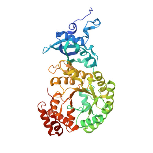
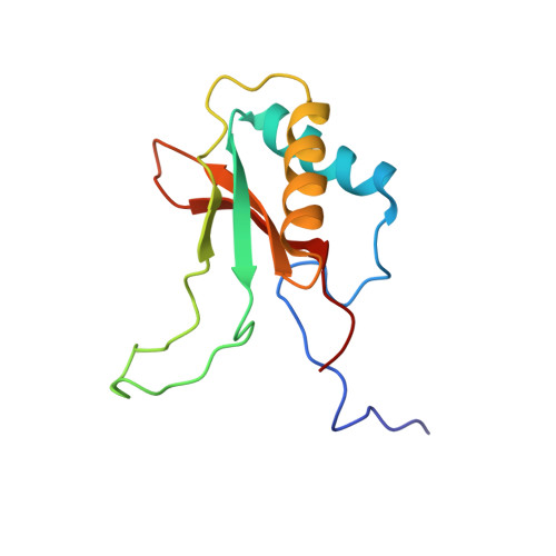
Structure of Pisum sativum Rubisco with bound ribulose 1,5-bisphosphate.

Loewen, P.C.Didychuk, A.L.Switala, J.Perez-Luque, R.Fita, I.Loewen, M.C.

(2013) Acta Crystallogr Sect F Struct Biol Cryst Commun 69: 10-14

  • DOI: https://doi.org/10.1107/S1744309112047549
  • Primary Citation of Related Structures:  
    4HHH

  • PubMed Abstract: 

    The first structure of a ribulose-1,5-bisphosphate carboxylase/oxygenase (Rubisco) from a pulse crop is reported. Rubisco was purified from Pisum sativum (garden pea) and diffraction-quality crystals were obtained by hanging-drop vapour diffusion in the presence of the substrate ribulose 1,5-bisphosphate. X-ray diffraction data were recorded to 2.20 Å resolution from a single crystal at the Canadian Light Source. The overall quaternary structure of non-activated P. sativum Rubisco highlights the conservation of the form I Rubisco hexadecameric complex. The electron density places the substrate in the active site at the interface of the large-subunit dimers. Lys201 in the active site is not carbamylated as expected for this non-activated structure. Some heterogeneity in the small-subunit sequence is noted, as well as possible variations in the conformation and contacts of ribulose 1,5-bisphosphate in the large-subunit active sites. Overall, the active-site conformation most closely correlates with the `closed' conformation observed in other substrate/inhibitor-bound Rubisco structures.


  • Organizational Affiliation

    Department of Microbiology, University of Manitoba, 418 Buller Building, Winnipeg, MB R3T 2N2, Canada.


Macromolecules
Find similar proteins by:  (by identity cutoff)  |  3D Structure
Entity ID: 1
MoleculeChains Sequence LengthOrganismDetailsImage
Ribulose bisphosphate carboxylase large chain
A, B, C, D
475Pisum sativumMutation(s): 0 
EC: 4.1.1.39
UniProt
Find proteins for P04717 (Pisum sativum)
Explore P04717 
Go to UniProtKB:  P04717
Entity Groups  
Sequence Clusters30% Identity50% Identity70% Identity90% Identity95% Identity100% Identity
UniProt GroupP04717
Sequence Annotations
Expand
  • Reference Sequence
Find similar proteins by:  (by identity cutoff)  |  3D Structure
Entity ID: 2
MoleculeChains Sequence LengthOrganismDetailsImage
Ribulose bisphosphate carboxylase small chainE [auth S],
F [auth T],
G [auth U],
H [auth V]
123Pisum sativumMutation(s): 0 
UniProt
Find proteins for P07689 (Pisum sativum)
Explore P07689 
Go to UniProtKB:  P07689
Entity Groups  
Sequence Clusters30% Identity50% Identity70% Identity90% Identity95% Identity100% Identity
UniProt GroupP07689
Sequence Annotations
Expand
  • Reference Sequence
Experimental Data & Validation

Experimental Data

  • Method: X-RAY DIFFRACTION
  • Resolution: 2.20 Å
  • R-Value Free: 0.279 
  • R-Value Work: 0.197 
  • R-Value Observed: 0.201 
  • Space Group: P 21 2 21
Unit Cell:
Length ( Å )Angle ( ˚ )
a = 109.79α = 90
b = 109.95β = 90
c = 201.44γ = 90
Software Package:
Software NamePurpose
SCALAdata scaling
REFMACrefinement
PDB_EXTRACTdata extraction
CLSIdata collection
MOSFLMdata reduction
PHENIXphasing

Structure Validation

View Full Validation Report


Ligand Structure Quality Assessment 


Entry History 

Deposition Data

Revision History  (Full details and data files)

  • Version 1.0: 2012-10-31
    Type: Initial release
  • Version 1.1: 2013-01-09
    Changes: Database references
  • Version 1.2: 2013-01-23
    Changes: Database references