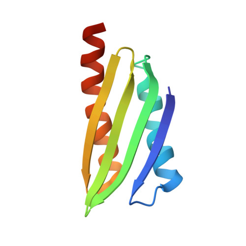
4HEI

2A X-RAY STRUCTURE OF HPF from VIBRIO CHOLERAE


Experimental Data Snapshot

  • Method: X-RAY DIFFRACTION
  • Resolution: 1.60 Å
  • R-Value Free: 0.281 
  • R-Value Work: 0.262 
  • R-Value Observed: 0.262 

wwPDB Validation   3D Report Full Report


This is version 1.6 of the entry. See complete history


Literature

Structure of Vibrio cholerae ribosome hibernation promoting factor.

De Bari, H.Berry, E.A.

(2013) Acta Crystallogr Sect F Struct Biol Cryst Commun 69: 228-236

  • DOI: https://doi.org/10.1107/S1744309113000961
  • Primary Citation of Related Structures:  
    4HEI

  • PubMed Abstract: 

    The X-ray crystal structure of ribosome hibernation promoting factor (HPF) from Vibrio cholerae is presented at 2.0 Å resolution. The crystal was phased by two-wavelength MAD using cocrystallized cobalt. The asymmetric unit contained two molecules of HPF linked by four Co atoms. The metal-binding sites observed in the crystal are probably not related to biological function. The structure of HPF has a typical β-α-β-β-β-α fold consistent with previous structures of YfiA and HPF from Escherichia coli. Comparison of the new structure with that of HPF from E. coli bound to the Thermus thermophilus ribosome [Polikanov et al. (2012), Science, 336, 915-918] shows that no significant structural changes are induced in HPF by binding.


  • Organizational Affiliation

    Biochemistry and Molecular Biology, SUNY Upstate Medical University, 750 E. Adams Avenue, Syracuse, NY 13210, USA.


Macromolecules
Find similar proteins by:  (by identity cutoff)  |  3D Structure
Entity ID: 1
MoleculeChains Sequence LengthOrganismDetailsImage
RIBOSOME HIBERNATION PROTEIN YHBH
A, B
95Vibrio cholerae O1 biovar El Tor str. N16961Mutation(s): 0 
UniProt
Find proteins for H9L4N9 (Vibrio cholerae serotype O1 (strain ATCC 39315 / El Tor Inaba N16961))
Explore H9L4N9 
Go to UniProtKB:  H9L4N9
Entity Groups  
Sequence Clusters30% Identity50% Identity70% Identity90% Identity95% Identity100% Identity
UniProt GroupH9L4N9
Sequence Annotations
Expand
  • Reference Sequence
Small Molecules
Ligands 3 Unique
IDChains Name / Formula / InChI Key2D Diagram3D Interactions
3CO
Query on 3CO

Download Ideal Coordinates CCD File 
C [auth A],
D [auth A],
K [auth B],
R [auth B]
COBALT (III) ION
Co
JAWGVVJVYSANRY-UHFFFAOYSA-N
SCN
Query on SCN

Download Ideal Coordinates CCD File 
E [auth A]
L [auth B]
M [auth B]
N [auth B]
P [auth B]
E [auth A],
L [auth B],
M [auth B],
N [auth B],
P [auth B],
Q [auth B]
THIOCYANATE ION
C N S
ZMZDMBWJUHKJPS-UHFFFAOYSA-M
UNL
Query on UNL

Download Ideal Coordinates CCD File 
F [auth A]
G [auth A]
H [auth A]
I [auth A]
J [auth A]
F [auth A],
G [auth A],
H [auth A],
I [auth A],
J [auth A],
O [auth B],
S [auth B]
Unknown ligand
ZMZDMBWJUHKJPS-UHFFFAOYSA-M
Experimental Data & Validation

Experimental Data

  • Method: X-RAY DIFFRACTION
  • Resolution: 1.60 Å
  • R-Value Free: 0.281 
  • R-Value Work: 0.262 
  • R-Value Observed: 0.262 
  • Space Group: P 41 21 2
Unit Cell:
Length ( Å )Angle ( ˚ )
a = 46.354α = 90
b = 46.354β = 90
c = 174.492γ = 90
Software Package:
Software NamePurpose
HKL-2000data collection
PHENIXmodel building
CNSrefinement
DENZOdata reduction
SCALEPACKdata scaling
PHENIXphasing

Structure Validation

View Full Validation Report


Entry History 

Deposition Data

Revision History  (Full details and data files)

  • Version 1.0: 2012-10-24
    Type: Initial release
  • Version 1.1: 2012-12-12
    Changes: Derived calculations
  • Version 1.2: 2013-03-06
    Changes: Database references, Derived calculations, Other
  • Version 1.3: 2013-05-22
    Changes: Database references
  • Version 1.4: 2017-06-21
    Changes: Database references, Source and taxonomy
  • Version 1.5: 2019-11-20
    Changes: Derived calculations
  • Version 1.6: 2024-02-28
    Changes: Data collection, Database references, Derived calculations