4GQS | pdb_00004gqs

Structure of Human Microsomal Cytochrome P450 (CYP) 2C19


Experimental Data Snapshot

  • Method: X-RAY DIFFRACTION
  • Resolution: 2.87 Å
  • R-Value Free: 
    0.296 (Depositor), 0.292 (DCC) 
  • R-Value Work: 
    0.250 (Depositor), 0.245 (DCC) 
  • R-Value Observed: 
    0.250 (Depositor) 

Starting Model: experimental
View more details

wwPDB Validation 3D Report Full Report

Validation slider image for 4GQS

Ligand Structure Quality Assessment 


This is version 1.2 of the entry. See complete history

Literature

Structural Characterization of Human Cytochrome P450 2C19: ACTIVE SITE DIFFERENCES BETWEEN P450s 2C8, 2C9, AND 2C19.

Reynald, R.L.Sansen, S.Stout, C.D.Johnson, E.F.

(2012) J Biological Chem 287: 44581-44591

  • DOI: https://doi.org/10.1074/jbc.M112.424895
  • Primary Citation Related Structures: 
    4GQS

  • PubMed Abstract: 

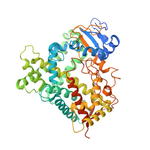
    To identify the structural features underlying the distinct substrate and inhibitor profiles of P450 2C19 relative to the closely related human enzymes, P450s 2C8 and 2C9, the atomic structure (Protein Data Bank code 4GQS) of cytochrome P450 2C19 complexed with the inhibitor (2-methyl-1-benzofuran-3-yl)-(4-hydroxy-3,5-dimethylphenyl)methanone (Protein Data Bank chemical component 0XV) was determined to 2.87 Å resolution by x-ray crystallography. The conformation of the peptide backbone of P450 2C19 is most similar to that of P450 2C8, but the substrate-binding cavity of P450 2C8 is much larger than that of P450 2C19 due to differences in the amino acid residues that form the substrate-binding cavities of the two enzymes. In contrast, the substrate-binding cavity of P450 2C19 is much more similar in size to that of the structure of the P450 2C9 flurbiprofen complex than to that of a modified P450 2C9 or that of P450 2C8. The cavities of the P450 2C19 0XV complex and the P450 2C9 flurbiprofen complex differ, however, because the helix B-C loops of the two enzymes are dissimilar. These conformational differences reflect the effects of adjacent structural elements that interact with the B-C loops and that differ between the two enzymes. The availability of a structure for 2C19 will facilitate computational approaches for predictions of substrate and inhibitor binding to this enzyme.


  • Organizational Affiliation
    • Department of Molecular and Experimental Medicine, The Scripps Research Institute, La Jolla, California 92037, USA.

Macromolecule Content 

  • Total Structure Weight: 222.19 kDa 
  • Atom Count: 15,043 
  • Modeled Residue Count: 1,834 
  • Deposited Residue Count: 1,908 
  • Unique protein chains: 1

Macromolecules

Find similar proteins by:|  3D Structure
Entity ID: 1
MoleculeChains  Sequence LengthOrganismDetailsImage
Cytochrome P450 2C19
A, B, C, D
477Homo sapiensMutation(s): 0 
Gene Names: CYP2C19
EC: 1.14.13 (PDB Primary Data), 1.14.13.80 (PDB Primary Data), 1.14.13.48 (PDB Primary Data), 1.14.13.49 (PDB Primary Data), 1.14.14.1 (UniProt), 1.14.14.75 (UniProt), 1.14.14.53 (UniProt), 1.14.14.52 (UniProt), 1.14.14.51 (UniProt)
Membrane Entity: Yes 
UniProt & NIH Common Fund Data Resources
Find proteins for P33261 (Homo sapiens)
Explore P33261 
Go to UniProtKB:  P33261
PHAROS:  P33261
GTEx:  ENSG00000165841 
Entity Groups
Sequence Clusters30% Identity50% Identity70% Identity90% Identity95% Identity100% Identity
UniProt GroupP33261
Sequence Annotations
Expand
Reference Sequence

Small Molecules

Ligands 3 Unique
IDChains Name / Formula / InChI Key2D Diagram3D Interactions
HEM

Query on HEM


Download:Ideal Coordinates CCD File
E [auth A],
J [auth B],
L [auth C],
N [auth D]
PROTOPORPHYRIN IX CONTAINING FE
C34 H32 Fe N4 O4
KABFMIBPWCXCRK-RGGAHWMASA-L
0XV

Query on 0XV


Download:Ideal Coordinates CCD File
F [auth A],
K [auth B],
M [auth C],
O [auth D]
(4-hydroxy-3,5-dimethylphenyl)(2-methyl-1-benzofuran-3-yl)methanone
C18 H16 O3
CIXADHNQIHMLIL-UHFFFAOYSA-N
GOL

Query on GOL


Download:Ideal Coordinates CCD File
G [auth A],
H [auth A],
I [auth A]
GLYCEROL
C3 H8 O3
PEDCQBHIVMGVHV-UHFFFAOYSA-N

Experimental Data & Validation

Experimental Data

  • Method: X-RAY DIFFRACTION
  • Resolution: 2.87 Å
  • R-Value Free:  0.296 (Depositor), 0.292 (DCC) 
  • R-Value Work:  0.250 (Depositor), 0.245 (DCC) 
  • R-Value Observed: 0.250 (Depositor) 
Space Group: H 3 2
Unit Cell:
Length ( Å )Angle ( ˚ )
a = 159.189α = 90
b = 159.189β = 90
c = 450.027γ = 120
Software Package:
Software NamePurpose
PHASERphasing
CNSrefinement
MOSFLMdata reduction
SCALAdata scaling

Structure Validation

View Full Validation Report


Ligand Structure Quality Assessment 


Entry History 

Deposition Data

Revision History  (Full details and data files)

  • Version 1.0: 2012-11-14
    Type: Initial release
  • Version 1.1: 2013-01-16
    Changes: Database references
  • Version 1.2: 2023-09-13
    Changes: Data collection, Database references, Derived calculations, Refinement description