Crystal Structure and Functional Analysis of JMJD5 Indicate an Alternate Specificity and Function.
Del Rizzo, P.A., Krishnan, S., Trievel, R.C.(2012) Mol Cell Biol 32: 4044-4052
- PubMed: 22851697 Search on PubMedSearch on PubMed Central
- DOI: https://doi.org/10.1128/MCB.00513-12
- Primary Citation Related Structures:
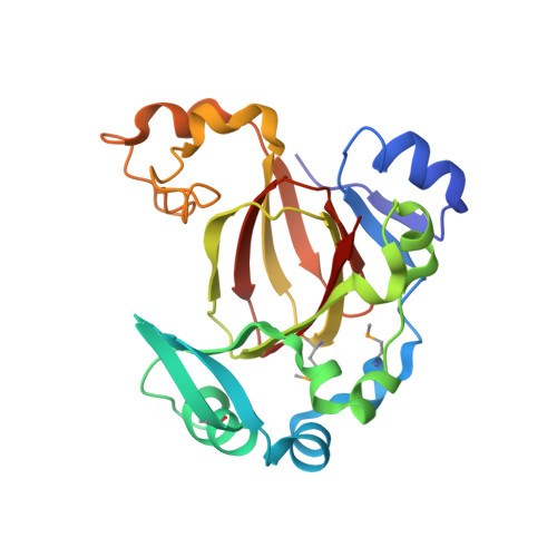
4GJY, 4GJZ - PubMed Abstract:
JMJD5 is a Jumonji C (JmjC) protein that has been implicated in breast cancer tumorigenesis, circadian rhythm regulation, embryological development, and osteoclastogenesis. Recently, JMJD5 (also called KDM8) has been reported to demethylate dimethylated Lys-36 in histone H3 (H3K36me2), regulating genes that control cell cycle progression. Here, we report high-resolution crystal structures of the human JMJD5 catalytic domain in complex with the substrate 2-oxoglutarate (2-OG) and the inhibitor N-oxalylglycine (NOG). The structures reveal a β-barrel fold that is conserved in the JmjC family and a long shallow cleft that opens into the enzyme's active site. A comparison with other JmjC enzymes illustrates that JMJD5 shares sequence and structural homology with the asparaginyl and histidinyl hydroxylase FIH-1 (factor inhibiting hypoxia-inducible factor 1 [HIF-1]), the lysyl hydroxylase JMJD6, and the RNA hydroxylase TYW5 but displays limited homology to JmjC lysine demethylases (KDMs). Contrary to previous findings, biochemical assays indicate that JMJD5 does not display demethylase activity toward methylated H3K36 nor toward the other methyllysines in the N-terminal tails of histones H3 and H4. Together, these results imply that JMJD5 participates in roles independent of histone demethylation and may function as a protein hydroxylase given its structural homology with FIH-1 and JMJD6.
- Department of Biological Chemistry, University of Michigan, Ann Arbor, MI, USA.
Organizational Affiliation: