4GH8 | pdb_00004gh8

Crystal structure of a 'humanized' E. coli dihydrofolate reductase


Experimental Data Snapshot

  • Method: X-RAY DIFFRACTION
  • Resolution: 1.85 Å
  • R-Value Free: 
    0.245 (Depositor), 0.250 (DCC) 
  • R-Value Work: 
    0.203 (Depositor), 0.210 (DCC) 
  • R-Value Observed: 
    0.205 (Depositor) 

wwPDB Validation   3D Report Full Report


Ligand Structure Quality Assessment 


This is version 1.3 of the entry. See complete history


Literature

Functional significance of evolving protein sequence in dihydrofolate reductase from bacteria to humans.

Liu, C.T.Hanoian, P.French, J.B.Pringle, T.H.Hammes-Schiffer, S.Benkovic, S.J.

(2013) Proc Natl Acad Sci U S A 110: 10159-10164

  • DOI: https://doi.org/10.1073/pnas.1307130110
  • Primary Citation of Related Structures:  
    4GH8

  • PubMed Abstract: 

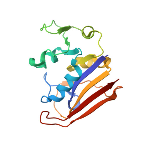
    With the rapidly growing wealth of genomic data, experimental inquiries on the functional significance of important divergence sites in protein evolution are becoming more accessible. Here we trace the evolution of dihydrofolate reductase (DHFR) and identify multiple key divergence sites among 233 species between humans and bacteria. We connect these sites, experimentally and computationally, to changes in the enzyme's binding properties and catalytic efficiency. One of the identified evolutionarily important sites is the N23PP modification (∼mid-Devonian, 415-385 Mya), which alters the conformational states of the active site loop in Escherichia coli dihydrofolate reductase and negatively impacts catalysis. This enzyme activity was restored with the inclusion of an evolutionarily significant lid domain (G51PEKN in E. coli enzyme; ∼2.4 Gya). Guided by this evolutionary genomic analysis, we generated a human-like E. coli dihydrofolate reductase variant through three simple mutations despite only 26% sequence identity between native human and E. coli DHFRs. Molecular dynamics simulations indicate that the overall conformational motions of the protein within a common scaffold are retained throughout evolution, although subtle changes to the equilibrium conformational sampling altered the free energy barrier of the enzymatic reaction in some cases. The data presented here provide a glimpse into the evolutionary trajectory of functional DHFR through its protein sequence space that lead to the diverged binding and catalytic properties of the E. coli and human enzymes.


  • Organizational Affiliation
    • Department of Chemistry, Pennsylvania State University, University Park, PA 16802, USA.

Macromolecules
Find similar proteins by:  (by identity cutoff)  |  3D Structure
Entity ID: 1
MoleculeChains Sequence LengthOrganismDetailsImage
Dihydrofolate reductaseA [auth B],
B [auth A]
162Escherichia coli K-12Mutation(s): 0 
Gene Names: folAtmrAb0048JW0047
EC: 1.5.1.3
UniProt
Find proteins for P0ABQ4 (Escherichia coli (strain K12))
Explore P0ABQ4 
Go to UniProtKB:  P0ABQ4
Entity Groups  
Sequence Clusters30% Identity50% Identity70% Identity90% Identity95% Identity100% Identity
UniProt GroupP0ABQ4
Sequence Annotations
Expand
  • Reference Sequence
Small Molecules
Ligands 3 Unique
IDChains Name / Formula / InChI Key2D Diagram3D Interactions
NDP
Query on NDP

Download Ideal Coordinates CCD File 
D [auth B],
G [auth A]
NADPH DIHYDRO-NICOTINAMIDE-ADENINE-DINUCLEOTIDE PHOSPHATE
C21 H30 N7 O17 P3
ACFIXJIJDZMPPO-NNYOXOHSSA-N
MTX
Query on MTX

Download Ideal Coordinates CCD File 
C [auth B],
F [auth A]
METHOTREXATE
C20 H22 N8 O5
FBOZXECLQNJBKD-ZDUSSCGKSA-N
CA
Query on CA

Download Ideal Coordinates CCD File 
E [auth B],
H [auth A],
I [auth A]
CALCIUM ION
Ca
BHPQYMZQTOCNFJ-UHFFFAOYSA-N
Binding Affinity Annotations 
IDSourceBinding Affinity
MTX BindingDB:  4GH8 IC50: min: 3, max: 8.8 (nM) from 3 assay(s)
EC50: 1 (nM) from 1 assay(s)
Experimental Data & Validation

Experimental Data

  • Method: X-RAY DIFFRACTION
  • Resolution: 1.85 Å
  • R-Value Free:  0.245 (Depositor), 0.250 (DCC) 
  • R-Value Work:  0.203 (Depositor), 0.210 (DCC) 
  • R-Value Observed: 0.205 (Depositor) 
Space Group: P 1 21 1
Unit Cell:
Length ( Å )Angle ( ˚ )
a = 52.245α = 90
b = 63.773β = 106.82
c = 62.439γ = 90
Software Package:
Software NamePurpose
DENZOdata reduction
SCALEPACKdata scaling
MOLREPphasing
REFMACrefinement
PDB_EXTRACTdata extraction
ADSCdata collection

Structure Validation

View Full Validation Report


Ligand Structure Quality Assessment 


Entry History 

Deposition Data

Revision History  (Full details and data files)

  • Version 1.0: 2013-06-05
    Type: Initial release
  • Version 1.1: 2013-07-03
    Changes: Database references
  • Version 1.2: 2017-11-15
    Changes: Refinement description
  • Version 1.3: 2024-02-28
    Changes: Data collection, Database references, Derived calculations