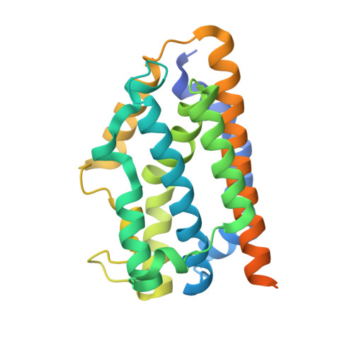
Discrimination between CO and O(2) in heme oxygenase: comparison of static structures and dynamic conformation changes following CO photolysis.
Sugishima, M., Moffat, K., Noguchi, M.(2012) Biochemistry 51: 8554-8562
- PubMed: 23043644 Search on PubMedSearch on PubMed Central
- DOI: https://doi.org/10.1021/bi301175x
- Primary Citation Related Structures:
4G7L, 4G7P, 4G7T, 4G7U, 4G8P, 4G8U, 4G8W, 4G98, 4G99 - PubMed Abstract:
Heme oxygenase (HO) catalyzes heme degradation, one of its products being carbon monoxide (CO). It is well known that CO has a higher affinity for heme iron than does molecular oxygen (O(2)); therefore, CO is potentially toxic. Because O(2) is required for the HO reaction, HO must discriminate effectively between CO and O(2) and thus escape product inhibition. Previously, we demonstrated large conformational changes in the heme-HO-1 complex upon CO binding that arise from steric hindrance between CO bound to the heme iron and Gly-139. However, we have not yet identified those changes that are specific to CO binding and do not occur upon O(2) binding. Here we determine the crystal structure of the O(2)-bound form at 1.8 Å resolution and reveal the structural changes that are specific to CO binding. Moreover, difference Fourier maps comparing the structures before and after CO photolysis at <160 K clearly show structural changes such as movement of the distal F-helix upon CO photolysis. No such changes are observed upon O(2) photolysis, consistent with the structures of the ligand-free, O(2)-bound, and CO-bound forms. Protein motions even at cryogenic temperatures imply that the CO-bound heme-HO-1 complex is severely constrained (as in ligand binding to the T-state of hemoglobin), indicating that CO binding to the heme-HO-1 complex is specifically inhibited by steric hindrance. The difference Fourier maps also suggest new routes for CO migration.
- Department of Medical Biochemistry, Kurume University School of Medicine, 67 Asahi-machi, Kurume 830-0011, Japan. sugishima_masakazu@med.kurume-u.ac.jp
Organizational Affiliation: