Comparative Analysis of Folding and Substrate Binding Sites between Regulated Hexameric Type II Citrate Synthases and Unregulated Dimeric Type I Enzymes.
Nguyen, N.T., Maurus, R., Stokell, D.J., Ayed, A., Duckworth, H.W., Brayer, G.D.(2001) Biochemistry 40: 13177
- PubMed: 11683626 Search on PubMed
- DOI: https://doi.org/10.1021/bi010408o
- Primary Citation Related Structures:
4G6B - PubMed Abstract:
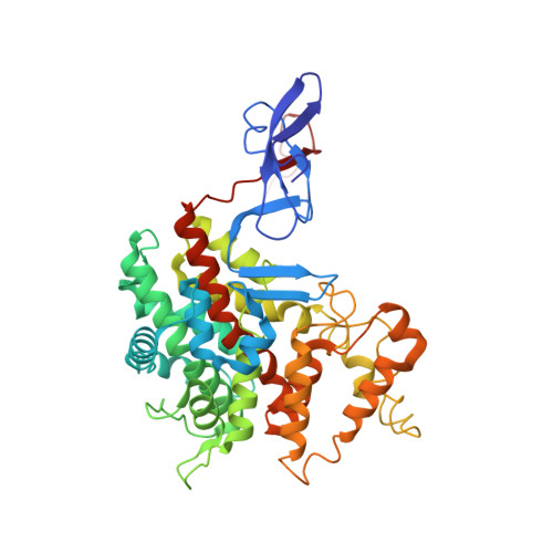
We describe the first structure determination of a type II citrate synthase, an enzyme uniquely found in Gram-negative bacteria. Such enzymes are hexameric and are strongly and specifically inhibited by NADH through an allosteric mechanism. This is in contrast to the widespread dimeric type I citrate synthases found in other organisms, which do not show allosteric properties. Our structure of the hexameric type II citrate synthase from Escherichia coli is composed of three identical dimer units arranged about a central 3-fold axis. The interactions that lead to hexamer formation are concentrated in a relatively small region composed of helix F, FG and IJ helical turns, and a seven-residue loop between helices J and K. This latter loop is present only in type II citrate synthase sequences. Running through the middle of the hexamer complex, and along the 3-fold axis relating dimer units, is a remarkable pore lined with 18 cationic residues and an associated hydrogen-bonded network. Also unexpected was the observation of a novel N-terminal domain, formed by the collective interactions of the first 52 residues from the two subunits of each dimer. The domain formed is rich in beta-sheet structure and has no counterpart in previous structural studies of type I citrate synthases. This domain is located well away from the dimer-dimer contacts that form the hexamer, and it is not involved in hexamer formation. Another surprising observation from the structure of type II E. coli citrate synthase is the unusual polypeptide chain folding found at the putative acetylcoenzyme A binding site. Key parts of this region, including His264 and a portion of polypeptide chain known from type I structures to form an adenine binding loop (residues 299-303), are shifted by as much as 10 A from where they must be for substrate binding and catalysis to occur. Furthermore, the adjacent polypeptide chain composed of residues 267-297 is extremely mobile in our structure. Thus, acetylcoenzyme A binding to type II E. coli citrate synthase would require substantial structural shifts and a concerted refolding of the polypeptide chain to form an appropriate binding subsite. We propose that this essential rearrangement of the acetylcoenzyme A binding part of the active site is also a major feature of allostery in type II citrate synthases. Overall, this study suggests that the evolutionary development of hexameric association, the elaboration of a novel N-terminal domain, introduction of a NADH binding site, and the need to refold a key substrate binding site are all elements that have been developed to allow for the allosteric control of catalysis in the type II citrate synthases.
- Department of Biochemistry and Molecular Biology, University of British Columbia, Vancouver V6T 1Z3, Canada.
Organizational Affiliation: