4G0R

Structural characterization of H-1 Parvovirus: comparison of infectious virions to replication defective particles


Experimental Data Snapshot

  • Method: X-RAY DIFFRACTION
  • Resolution: 2.70 Å
  • R-Value Free: 0.217 
  • R-Value Work: 0.216 

wwPDB Validation   3D Report Full Report


Ligand Structure Quality Assessment 


This is version 1.2 of the entry. See complete history


Literature

Structural characterization of h-1 parvovirus: comparison of infectious virions to empty capsids.

Halder, S.Nam, H.J.Govindasamy, L.Vogel, M.Dinsart, C.Salome, N.McKenna, R.Agbandje-McKenna, M.

(2013) J Virol 87: 5128-5140

  • DOI: https://doi.org/10.1128/JVI.03416-12
  • Primary Citation of Related Structures:  
    4G0R, 4GBT

  • PubMed Abstract: 

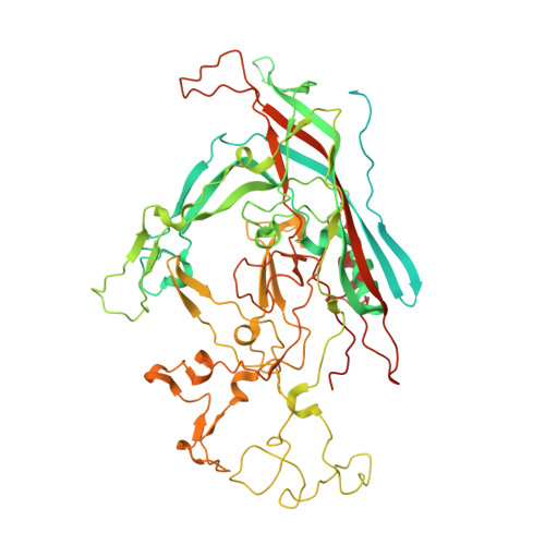
    The structure of single-stranded DNA (ssDNA) packaging H-1 parvovirus (H-1PV), which is being developed as an antitumor gene delivery vector, has been determined for wild-type (wt) virions and noninfectious (empty) capsids to 2.7- and 3.2-Å resolution, respectively, using X-ray crystallography. The capsid viral protein (VP) structure consists of an α-helix and an eight-stranded anti-parallel β-barrel with large loop regions between the strands. The β-barrel and loops form the capsid core and surface, respectively. In the wt structure, 600 nucleotides are ordered in an interior DNA binding pocket of the capsid. This accounts for ∼12% of the H-1PV genome. The wt structure is identical to the empty capsid structure, except for side chain conformation variations at the nucleotide binding pocket. Comparison of the H-1PV nucleotides to those observed in canine parvovirus and minute virus of mice, two members of the genus Parvovirus, showed both similarity in structure and analogous interactions. This observation suggests a functional role, such as in capsid stability and/or ssDNA genome recognition for encapsulation. The VP structure differs from those of other parvoviruses in surface loop regions that control receptor binding, tissue tropism, pathogenicity, and antibody recognition, including VP sequences reported to determine tumor cell tropism for oncotropic rodent parvoviruses. These structures of H-1PV provide insight into structural features that dictate capsid stabilization following genome packaging and three-dimensional information applicable for rational design of tumor-targeted recombinant gene delivery vectors.


  • Organizational Affiliation

    Department of Biochemistry and Molecular Biology, University of Florida, Gainesville, Florida, USA.


Macromolecules

Find similar proteins by:  (by identity cutoff)  |  3D Structure
Entity ID: 1
MoleculeChains Sequence LengthOrganismDetailsImage
Capsid protein VP1735H-1 parvovirusMutation(s): 0 
UniProt
Find proteins for P03136 (Hamster parvovirus H1)
Explore P03136 
Go to UniProtKB:  P03136
Entity Groups  
Sequence Clusters30% Identity50% Identity70% Identity90% Identity95% Identity100% Identity
UniProt GroupP03136
Sequence Annotations
Expand
  • Reference Sequence

Find similar nucleic acids by:  Sequence   |   3D Structure  

Entity ID: 2
MoleculeChains LengthOrganismImage
DNA (5'-D(P*CP*TP*GP*AP*CP*TP*TP*CP*AP*A)-3')B [auth C]10H-1 parvovirus
Sequence Annotations
Expand
  • Reference Sequence
Small Molecules
Ligands 5 Unique
IDChains Name / Formula / InChI Key2D Diagram3D Interactions
DC
Query on DC

Download Ideal Coordinates CCD File 
F [auth A]2'-DEOXYCYTIDINE-5'-MONOPHOSPHATE
C9 H14 N3 O7 P
NCMVOABPESMRCP-SHYZEUOFSA-N
EDO
Query on EDO

Download Ideal Coordinates CCD File 
C [auth A],
D [auth A],
E [auth A],
H [auth A]
1,2-ETHANEDIOL
C2 H6 O2
LYCAIKOWRPUZTN-UHFFFAOYSA-N
CL
Query on CL

Download Ideal Coordinates CCD File 
J [auth A]CHLORIDE ION
Cl
VEXZGXHMUGYJMC-UHFFFAOYSA-M
MG
Query on MG

Download Ideal Coordinates CCD File 
K [auth C],
L [auth C]
MAGNESIUM ION
Mg
JLVVSXFLKOJNIY-UHFFFAOYSA-N
NA
Query on NA

Download Ideal Coordinates CCD File 
G [auth A],
I [auth A]
SODIUM ION
Na
FKNQFGJONOIPTF-UHFFFAOYSA-N
Experimental Data & Validation

Experimental Data

  • Method: X-RAY DIFFRACTION
  • Resolution: 2.70 Å
  • R-Value Free: 0.217 
  • R-Value Work: 0.216 
  • Space Group: P 1 21 1
Unit Cell:
Length ( Å )Angle ( ˚ )
a = 255.4α = 90
b = 350.4β = 90.34
c = 271.6γ = 90
Software Package:
Software NamePurpose
HKL-2000data collection
AMoREphasing
CNSrefinement
HKL-2000data reduction
SCALEPACKdata scaling

Structure Validation

View Full Validation Report


Ligand Structure Quality Assessment 


Entry History 

Deposition Data

Revision History  (Full details and data files)

  • Version 1.0: 2013-02-27
    Type: Initial release
  • Version 1.1: 2013-04-24
    Changes: Database references
  • Version 1.2: 2024-02-28
    Changes: Data collection, Database references, Derived calculations