4FDJ

The molecular basis of mucopolysaccharidosis IV A, complex with GalNAc


Experimental Data Snapshot

  • Method: X-RAY DIFFRACTION
  • Resolution: 2.81 Å
  • R-Value Free: 0.251 
  • R-Value Work: 0.207 
  • R-Value Observed: 0.209 

wwPDB Validation   3D Report Full Report


Ligand Structure Quality Assessment 


This is version 2.2 of the entry. See complete history


Literature

The Structure of Human GALNS Reveals the Molecular Basis for Mucopolysaccharidosis IV A.

Rivera-Colon, Y.Schutsky, E.K.Kita, A.Z.Garman, S.C.

(2012) J Mol Biol 423: 736-751

  • DOI: https://doi.org/10.1016/j.jmb.2012.08.020
  • Primary Citation of Related Structures:  
    4FDI, 4FDJ

  • PubMed Abstract: 

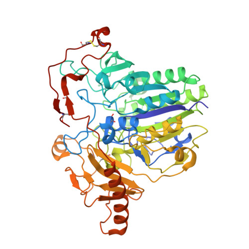
    Lysosomal enzymes catalyze the breakdown of macromolecules in the cell. In humans, loss of activity of a lysosomal enzyme leads to an inherited metabolic defect known as a lysosomal storage disorder. The human lysosomal enzyme galactosamine-6-sulfatase (GALNS, also known as N-acetylgalactosamine-6-sulfatase and GalN6S; E.C. 3.1.6.4) is deficient in patients with the lysosomal storage disease mucopolysaccharidosis IV A (also known as MPS IV A and Morquio A). Here, we report the three-dimensional structure of human GALNS, determined by X-ray crystallography at 2.2Å resolution. The structure reveals a catalytic gem diol nucleophile derived from modification of a cysteine side chain. The active site of GALNS is a large, positively charged trench suitable for binding polyanionic substrates such as keratan sulfate and chondroitin-6-sulfate. Enzymatic assays on the insect-cell-expressed human GALNS indicate activity against synthetic substrates and inhibition by both substrate and product. Mapping 120 MPS IV A missense mutations onto the structure reveals that a majority of mutations affect the hydrophobic core of the structure, indicating that most MPS IV A cases result from misfolding of GALNS. Comparison of the structure of GALNS to paralogous sulfatases shows a wide variety of active-site geometries in the family but strict conservation of the catalytic machinery. Overall, the structure and the known mutations establish the molecular basis for MPS IV A and for the larger MPS family of diseases.


  • Organizational Affiliation

    Department of Biochemistry and Molecular Biology, University of Massachusetts Amherst, Amherst, MA 01003, USA.


Macromolecules
Find similar proteins by:  (by identity cutoff)  |  3D Structure
Entity ID: 1
MoleculeChains Sequence LengthOrganismDetailsImage
N-acetylgalactosamine-6-sulfatase
A, B
502Homo sapiensMutation(s): 0 
Gene Names: GALNS
EC: 3.1.6.4
UniProt & NIH Common Fund Data Resources
Find proteins for P34059 (Homo sapiens)
Explore P34059 
Go to UniProtKB:  P34059
PHAROS:  P34059
GTEx:  ENSG00000141012 
Entity Groups  
Sequence Clusters30% Identity50% Identity70% Identity90% Identity95% Identity100% Identity
UniProt GroupP34059
Sequence Annotations
Expand
  • Reference Sequence
Small Molecules
Ligands 3 Unique
IDChains Name / Formula / InChI Key2D Diagram3D Interactions
NGA
Query on NGA

Download Ideal Coordinates CCD File 
F [auth A],
J [auth B]
2-acetamido-2-deoxy-beta-D-galactopyranose
C8 H15 N O6
OVRNDRQMDRJTHS-JAJWTYFOSA-N
NAG
Query on NAG

Download Ideal Coordinates CCD File 
C [auth A],
D [auth A],
G [auth B],
H [auth B]
2-acetamido-2-deoxy-beta-D-glucopyranose
C8 H15 N O6
OVRNDRQMDRJTHS-FMDGEEDCSA-N
CA
Query on CA

Download Ideal Coordinates CCD File 
E [auth A],
I [auth B]
CALCIUM ION
Ca
BHPQYMZQTOCNFJ-UHFFFAOYSA-N
Modified Residues  1 Unique
IDChains TypeFormula2D DiagramParent
DDZ
Query on DDZ
A, B
L-PEPTIDE LINKINGC3 H7 N O4ALA
Experimental Data & Validation

Experimental Data

  • Method: X-RAY DIFFRACTION
  • Resolution: 2.81 Å
  • R-Value Free: 0.251 
  • R-Value Work: 0.207 
  • R-Value Observed: 0.209 
  • Space Group: P 1 21 1
Unit Cell:
Length ( Å )Angle ( ˚ )
a = 60.706α = 90
b = 155.835β = 113.43
c = 61.801γ = 90
Software Package:
Software NamePurpose
SCALEPACKdata scaling
REFMACrefinement
PDB_EXTRACTdata extraction
HKL-2000data collection
HKL-2000data reduction
HKL-2000data scaling
REFMACphasing

Structure Validation

View Full Validation Report


Ligand Structure Quality Assessment 


Entry History 

Deposition Data

Revision History  (Full details and data files)

  • Version 1.0: 2012-09-05
    Type: Initial release
  • Version 1.1: 2012-09-19
    Changes: Database references
  • Version 1.2: 2012-10-31
    Changes: Database references
  • Version 2.0: 2020-07-29
    Type: Remediation
    Reason: Carbohydrate remediation
    Changes: Data collection, Database references, Derived calculations, Polymer sequence, Structure summary
  • Version 2.1: 2023-09-13
    Changes: Data collection, Database references, Refinement description, Structure summary
  • Version 2.2: 2023-12-06
    Changes: Data collection, Derived calculations