4EKS | pdb_00004eks

T4 Lysozyme L99A/M102H with Isoxazole Bound


Experimental Data Snapshot

  • Method: X-RAY DIFFRACTION
  • Resolution: 1.64 Å
  • R-Value Free: 
    0.198 (Depositor), 0.195 (DCC) 
  • R-Value Work: 
    0.171 (Depositor), 0.168 (DCC) 
  • R-Value Observed: 
    0.172 (Depositor) 

Starting Model: experimental
View more details

wwPDB Validation 3D Report Full Report

Validation slider image for 4EKS

Ligand Structure Quality Assessment 


This is version 1.3 of the entry. See complete history

Literature

Engineering a model protein cavity to catalyze the Kemp elimination.

Merski, M.Shoichet, B.K.

(2012) Proc Natl Acad Sci U S A 109: 16179-16183

  • DOI: https://doi.org/10.1073/pnas.1208076109
  • Primary Citation Related Structures: 
    4E97, 4EKP, 4EKQ, 4EKR, 4EKS

  • PubMed Abstract: 

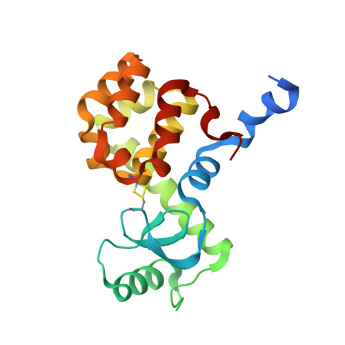
    Synthetic cavitands and protein cavities have been widely studied as models for ligand recognition. Here we investigate the Met102 → His substitution in the artificial L99A cavity in T4 lysozyme as a Kemp eliminase. The resulting enzyme had k(cat)/K(M) = 0.43 M(-1) s(-1) and a (k(cat)/K(M))/k(uncat) = 10(7) at pH 5.0. The crystal structure of this enzyme was determined at 1.30 Å, as were the structures of four complexes of substrate and product analogs. The absence of ordered waters or hydrogen bonding interactions, and the presence of a common catalytic base (His102) in an otherwise hydrophobic, buried cavity, facilitated detailed analysis of the reaction mechanism and its optimization. Subsequent substitutions increased eliminase activity by an additional four-fold. As activity-enhancing substitutions were engineered into the cavity, protein stability decreased, consistent with the stability-function trade-off hypothesis. This and related model cavities may provide templates for studying protein design principles in radically simplified environments.


  • Organizational Affiliation
    • Department of Pharmaceutical Chemistry, University of California-San Francisco, San Francisco, CA 94158-2550, USA.

Macromolecule Content 

  • Total Structure Weight: 44.17 kDa 
  • Atom Count: 3,252 
  • Modeled Residue Count: 349 
  • Deposited Residue Count: 374 
  • Unique protein chains: 1

Macromolecules

Find similar proteins by:|  3D Structure
Entity ID: 1
MoleculeChains  Sequence LengthOrganismDetailsImage
Lysozyme
A, B
187Tequatrovirus T4Mutation(s): 8 
Gene Names: E
EC: 3.2.1.17
UniProt
Find proteins for P00720 (Enterobacteria phage T4)
Explore P00720 
Go to UniProtKB:  P00720
Entity Groups
Sequence Clusters30% Identity50% Identity70% Identity90% Identity95% Identity100% Identity
UniProt GroupP00720
Sequence Annotations
Expand
Reference Sequence

Small Molecules

Ligands 5 Unique
IDChains Name / Formula / InChI Key2D Diagram3D Interactions
HED

Query on HED


Download:Ideal Coordinates CCD File
Q [auth B]2-HYDROXYETHYL DISULFIDE
C4 H10 O2 S2
KYNFOMQIXZUKRK-UHFFFAOYSA-N
0R1

Query on 0R1


Download:Ideal Coordinates CCD File
D [auth A],
L [auth B]
1,2-benzisoxazole
C7 H5 N O
KTZQTRPPVKQPFO-UHFFFAOYSA-N
SO4

Query on SO4


Download:Ideal Coordinates CCD File
E [auth A],
F [auth A],
G [auth A],
M [auth B],
N [auth B]
SULFATE ION
O4 S
QAOWNCQODCNURD-UHFFFAOYSA-L
BME

Query on BME


Download:Ideal Coordinates CCD File
C [auth A],
I [auth A],
K [auth B],
P [auth B]
BETA-MERCAPTOETHANOL
C2 H6 O S
DGVVWUTYPXICAM-UHFFFAOYSA-N
ACT

Query on ACT


Download:Ideal Coordinates CCD File
H [auth A],
J [auth A],
O [auth B]
ACETATE ION
C2 H3 O2
QTBSBXVTEAMEQO-UHFFFAOYSA-M

Experimental Data & Validation

Experimental Data

  • Method: X-RAY DIFFRACTION
  • Resolution: 1.64 Å
  • R-Value Free:  0.198 (Depositor), 0.195 (DCC) 
  • R-Value Work:  0.171 (Depositor), 0.168 (DCC) 
  • R-Value Observed: 0.172 (Depositor) 
Space Group: P 1 21 1
Unit Cell:
Length ( Å )Angle ( ˚ )
a = 48.3α = 90
b = 75.37β = 93.15
c = 52.65γ = 90
Software Package:
Software NamePurpose
XSCALEdata scaling
PHENIXrefinement
PDB_EXTRACTdata extraction
Blu-Icedata collection
XDSdata reduction
PHENIXphasing

Structure Validation

View Full Validation Report


Ligand Structure Quality Assessment 


Entry History 

Deposition Data

Revision History  (Full details and data files)

  • Version 1.0: 2012-09-05
    Type: Initial release
  • Version 1.1: 2012-10-03
    Changes: Database references
  • Version 1.2: 2012-10-17
    Changes: Database references
  • Version 1.3: 2023-09-13
    Changes: Data collection, Database references, Derived calculations, Refinement description