4EKK

Akt1 with AMP-PNP


Experimental Data Snapshot

  • Method: X-RAY DIFFRACTION
  • Resolution: 2.80 Å
  • R-Value Free: 0.280 
  • R-Value Work: 0.217 
  • R-Value Observed: 0.220 

wwPDB Validation   3D Report Full Report


This is version 1.1 of the entry. See complete history


Literature

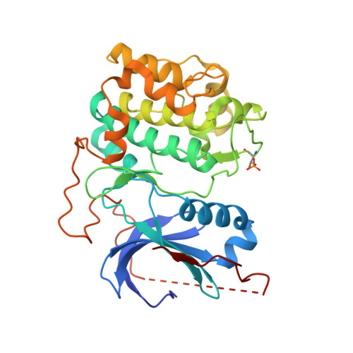
An ATP-Site On-Off Switch That Restricts Phosphatase Accessibility of Akt.

Lin, K.Lin, J.Wu, W.I.Ballard, J.Lee, B.B.Gloor, S.L.Vigers, G.P.Morales, T.H.Friedman, L.S.Skelton, N.Brandhuber, B.J.

(2012) Sci Signal 5: ra37-ra37

  • DOI: https://doi.org/10.1126/scisignal.2002618
  • Primary Citation of Related Structures:  
    4EKK, 4EKL

  • PubMed Abstract: 

    The protein serine-threonine kinase Akt undergoes a substantial conformational change upon activation, which is induced by the phosphorylation of two critical regulatory residues, threonine 308 and serine 473. Paradoxically, treating cells with adenosine 5'-triphosphate (ATP)-competitive inhibitors of Akt results in increased phosphorylation of both residues. We show that binding of ATP-competitive inhibitors stabilized a conformation in which both phosphorylated sites were inaccessible to phosphatases. ATP binding also produced this protection of the phosphorylated sites, whereas interaction with its hydrolysis product adenosine 5'-diphosphate (ADP) or allosteric Akt inhibitors resulted in increased accessibility of these phosphorylated residues. ATP-competitive inhibitors mimicked ATP by targeting active Akt. Forms of Akt activated by an oncogenic mutation or myristoylation were more potently inhibited by the ATP-competitive inhibitors than was wild-type Akt. These data support a new model of kinase regulation, wherein nucleotides modulate an on-off switch in Akt through conformational changes, which is disrupted by ATP-competitive inhibitors.


  • Organizational Affiliation

    Genentech Inc., South San Francisco, CA 94080, USA. klin@gene.com


Macromolecules
Find similar proteins by:  (by identity cutoff)  |  3D Structure
Entity ID: 1
MoleculeChains Sequence LengthOrganismDetailsImage
RAC-alpha serine/threonine-protein kinase
A, B
341Homo sapiensMutation(s): 1 
Gene Names: AKT1PKBRAC
EC: 2.7.11.1
UniProt & NIH Common Fund Data Resources
Find proteins for P31749 (Homo sapiens)
Explore P31749 
Go to UniProtKB:  P31749
PHAROS:  P31749
Entity Groups  
Sequence Clusters30% Identity50% Identity70% Identity90% Identity95% Identity100% Identity
UniProt GroupP31749
Sequence Annotations
Expand
  • Reference Sequence

Find similar proteins by:  Sequence   |   3D Structure  

Entity ID: 2
MoleculeChains Sequence LengthOrganismDetailsImage
Glycogen synthase kinase-3 beta
C, D
10Homo sapiensMutation(s): 0 
Gene Names: GSK3B
EC: 2.7.11.26 (PDB Primary Data), 2.7.11.1 (PDB Primary Data)
UniProt & NIH Common Fund Data Resources
Find proteins for P49841 (Homo sapiens)
Explore P49841 
Go to UniProtKB:  P49841
PHAROS:  P49841
Entity Groups  
Sequence Clusters30% Identity50% Identity70% Identity90% Identity95% Identity100% Identity
UniProt GroupP49841
Sequence Annotations
Expand
  • Reference Sequence
Experimental Data & Validation

Experimental Data

  • Method: X-RAY DIFFRACTION
  • Resolution: 2.80 Å
  • R-Value Free: 0.280 
  • R-Value Work: 0.217 
  • R-Value Observed: 0.220 
  • Space Group: P 1 21 1
Unit Cell:
Length ( Å )Angle ( ˚ )
a = 86.637α = 90
b = 55.853β = 103.43
c = 91.402γ = 90
Software Package:
Software NamePurpose
CrystalCleardata collection
MOLREPphasing
REFMACrefinement
MOSFLMdata reduction
SCALAdata scaling

Structure Validation

View Full Validation Report


Entry History 

Deposition Data

Revision History  (Full details and data files)

  • Version 1.0: 2012-05-23
    Type: Initial release
  • Version 1.1: 2018-01-24
    Changes: Structure summary