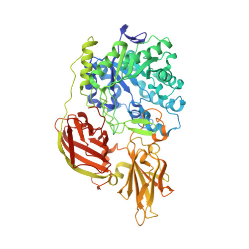
4E8C

Crystal structure of streptococcal beta-galactosidase in complex with galactose


Experimental Data Snapshot

  • Method: X-RAY DIFFRACTION
  • Resolution: 1.95 Å
  • R-Value Free: 0.243 
  • R-Value Work: 0.205 
  • R-Value Observed: 0.207 

Starting Model: experimental
View more details

wwPDB Validation   3D Report Full Report


Ligand Structure Quality Assessment 


This is version 1.4 of the entry. See complete history


Literature

Structural insights into the substrate specificity of Streptococcus pneumoniae beta (1,3)-galactosidase BgaC

Cheng, W.Wang, L.Jiang, Y.L.Bai, X.H.Chu, J.Li, Q.Yu, G.Liang, Q.L.Zhou, C.Z.Chen, Y.X.

(2012) J Biol Chem 287: 22910-22918

  • DOI: https://doi.org/10.1074/jbc.M112.367128
  • Primary Citation of Related Structures:  
    4E8C, 4E8D

  • PubMed Abstract: 

    The surface-exposed β-galactosidase BgaC from Streptococcus pneumoniae was reported to be a virulence factor because of its specific hydrolysis activity toward the β(1,3)-linked galactose and N-acetylglucosamine (Galβ(1,3)NAG) moiety of oligosaccharides on the host molecules. Here we report the crystal structure of BgaC at 1.8 Å and its complex with galactose at 1.95 Å. At pH 5.5-8.0, BgaC exists as a stable homodimer, each subunit of which consists of three distinct domains: a catalytic domain of a classic (β/α)(8) TIM barrel, followed by two all-β domains (ABDs) of unknown function. The side walls of the TIM β-barrel and a loop extended from the first ABD constitute the active site. Superposition of the galactose-complexed structure to the apo-form revealed significant conformational changes of residues Trp-243 and Tyr-455. Simulation of a putative substrate entrance tunnel and modeling of a complex structure with Galβ(1,3)NAG enabled us to assign three key residues to the specific catalysis. Site-directed mutagenesis in combination with activity assays further proved that residues Trp-240 and Tyr-455 contribute to stabilizing the N-acetylglucosamine moiety, whereas Trp-243 is critical for fixing the galactose ring. Moreover, we propose that BgaC and other galactosidases in the GH-35 family share a common domain organization and a conserved substrate-determinant aromatic residue protruding from the second domain.


  • Organizational Affiliation

    Hefei National Laboratory for Physical Sciences at Microscale and School of Life Sciences, University of Science and Technology of China, Hefei, Anhui 230026, China.


Macromolecules
Find similar proteins by:  (by identity cutoff)  |  3D Structure
Entity ID: 1
MoleculeChains Sequence LengthOrganismDetailsImage
Glycosyl hydrolase, family 35
A, B
595Streptococcus pneumoniae TIGR4Mutation(s): 0 
Gene Names: SP_0060
EC: 3.2.1.23
UniProt
Find proteins for A0A0H2UN19 (Streptococcus pneumoniae serotype 4 (strain ATCC BAA-334 / TIGR4))
Explore A0A0H2UN19 
Go to UniProtKB:  A0A0H2UN19
Entity Groups  
Sequence Clusters30% Identity50% Identity70% Identity90% Identity95% Identity100% Identity
UniProt GroupA0A0H2UN19
Sequence Annotations
Expand
  • Reference Sequence
Small Molecules
Ligands 2 Unique
IDChains Name / Formula / InChI Key2D Diagram3D Interactions
GAL
Query on GAL

Download Ideal Coordinates CCD File 
C [auth A],
P [auth B]
beta-D-galactopyranose
C6 H12 O6
WQZGKKKJIJFFOK-FPRJBGLDSA-N
GOL
Query on GOL

Download Ideal Coordinates CCD File 
D [auth A]
E [auth A]
F [auth A]
G [auth A]
H [auth A]
D [auth A],
E [auth A],
F [auth A],
G [auth A],
H [auth A],
I [auth A],
J [auth A],
K [auth A],
L [auth A],
M [auth A],
N [auth A],
O [auth A],
Q [auth B],
R [auth B],
S [auth B]
GLYCEROL
C3 H8 O3
PEDCQBHIVMGVHV-UHFFFAOYSA-N
Experimental Data & Validation

Experimental Data

  • Method: X-RAY DIFFRACTION
  • Resolution: 1.95 Å
  • R-Value Free: 0.243 
  • R-Value Work: 0.205 
  • R-Value Observed: 0.207 
  • Space Group: P 1 21 1
Unit Cell:
Length ( Å )Angle ( ˚ )
a = 80.351α = 90
b = 82.374β = 106.84
c = 99.596γ = 90
Software Package:
Software NamePurpose
ADSCdata collection
AMoREphasing
REFMACrefinement
HKL-2000data reduction
SCALAdata scaling

Structure Validation

View Full Validation Report


Ligand Structure Quality Assessment 


Entry History 

Deposition Data

Revision History  (Full details and data files)

  • Version 1.0: 2012-05-30
    Type: Initial release
  • Version 1.1: 2012-06-13
    Changes: Database references
  • Version 1.2: 2013-08-07
    Changes: Database references
  • Version 1.3: 2020-07-29
    Type: Remediation
    Reason: Carbohydrate remediation
    Changes: Data collection, Derived calculations, Structure summary
  • Version 1.4: 2023-11-08
    Changes: Data collection, Database references, Refinement description, Structure summary