Structural Characterization of Closely Related O-antigen Lipopolysaccharide (LPS) Chain Length Regulators.
Kalynych, S., Yao, D., Magee, J., Cygler, M.(2012) J Biological Chem 287: 15696-15705
- PubMed: 22437828 Search on PubMedSearch on PubMed Central
- DOI: https://doi.org/10.1074/jbc.M112.354837
- Primary Citation Related Structures:
4E29, 4E2C, 4E2H, 4E2L - PubMed Abstract:
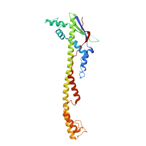
The surface O-antigen polymers of gram-negative bacteria exhibit a modal length distribution that depends on dedicated chain length regulator periplasmic proteins (polysaccharide co-polymerases, PCPs) anchored in the inner membrane by two transmembrane helices. In an attempt to determine whether structural changes underlie the O-antigen modal length specification, we have determined the crystal structures of several closely related PCPs, namely two chimeric PCP-1 family members solved at 1.6 and 2.8 Å and a wild-type PCP-1 from Shigella flexneri solved at 2.8 Å. The chimeric proteins form circular octamers, whereas the wild-type WzzB from S. flexneri was found to be an open trimer. We also present the structure of a Wzz(FepE) mutant, which exhibits severe attenuation in its ability to produce very long O-antigen polymers. Our findings suggest that the differences in the modal length distribution depend primarily on the surface-exposed amino acids in specific regions rather than on the differences in the oligomeric state of the PCP protomers.
- Department of Biochemistry, McGill University, Montreal, Quebec H3G 0B1, Canada.
Organizational Affiliation: