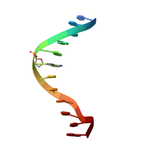
Methylation of the EcoRI recognition site does not alter DNA conformation: the crystal structure of d(CGCGAm6ATTCGCG) at 2.0-A resolution.
Frederick, C.A., Quigley, G.J., van der Marel, G.A., van Boom, J.H., Wang, A.H., Rich, A.(1988) J Biological Chem 263: 17872-17879
- PubMed: 2846582 Search on PubMed
- DOI: https://doi.org/10.2210/pdb4dnb/pdb
- Primary Citation Related Structures:
4DNB - PubMed Abstract:
Methylation of nucleic acid bases is known to prevent the cleavage of DNA by restriction endonucleases. The effect on the conformation of the DNA molecule itself and hence its interactions with other DNA binding proteins has been a subject of general interest. To help address this question, we have solved the crystal structure at 2.0 A of the methylated dodecamer, d(CGCGAm6ATTCGCG), which contains the EcoRI recognition sequence and have compared the conformation of the methylated molecule with that of its nonmethylated counterpart. This methylation produces a bulky hydrophobic patch on the floor of the major groove of B-DNA which plays an important role in the mechanism of inhibition of EcoRI restriction activity. However, with the exception of small perturbations in the immediate vicinity of the methyl groups, the structure is virtually unchanged. Given the lack of a conformational change upon methylation, we have extended this thesis of the recognition process to other types of restriction systems and found that different restriction enzymes seem to have their own characteristic protein-DNA interactions. The relative spatial orientations of methylation sites and cleavage sites must play a major role in ordering protein secondary structure elements as well as subunit-subunit interactions along the DNA strand.
- Department of Biology, Massachusetts Institute of Technology, Cambridge 02139.
Organizational Affiliation: