4DEX

Crystal structure of the Voltage Dependent Calcium Channel beta-2 Subunit in Complex With The CaV2.2 I-II Linker.


Experimental Data Snapshot

  • Method: X-RAY DIFFRACTION
  • Resolution: 2.00 Å
  • R-Value Free: 0.238 
  • R-Value Work: 0.198 
  • R-Value Observed: 0.200 

wwPDB Validation   3D Report Full Report


This is version 1.2 of the entry. See complete history


Literature

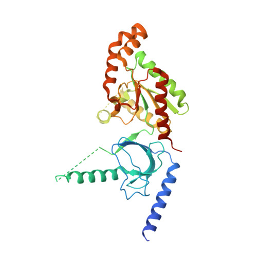
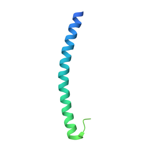
The role of a voltage-dependent Ca2+ channel intracellular linker: a structure-function analysis.

Almagor, L.Chomsky-Hecht, O.Ben-Mocha, A.Hendin-Barak, D.Dascal, N.Hirsch, J.A.

(2012) J Neurosci 32: 7602-7613

  • DOI: https://doi.org/10.1523/JNEUROSCI.5727-11.2012
  • Primary Citation of Related Structures:  
    4DEX, 4DEY

  • PubMed Abstract: 

    Voltage-dependent calcium channels (VDCCs) allow the passage of Ca(2+) ions through cellular membranes in response to membrane depolarization. The channel pore-forming subunit, α1, and a regulatory subunit (Ca(V)β) form a high affinity complex where Ca(V)β binds to a α1 interacting domain in the intracellular linker between α1 membrane domains I and II (I-II linker). We determined crystal structures of Ca(V)β2 functional core in complex with the Ca(V)1.2 and Ca(V)2.2 I-II linkers to a resolution of 1.95 and 2.0 Å, respectively. Structural differences between the highly conserved linkers, important for coupling Ca(V)β to the channel pore, guided mechanistic functional studies. Electrophysiological measurements point to the importance of differing linker structure in both Ca(V)1 and 2 subtypes with mutations affecting both voltage- and calcium-dependent inactivation and voltage dependence of activation. These linker effects persist in the absence of Ca(V)β, pointing to the intrinsic role of the linker in VDCC function and suggesting that I-II linker structure can serve as a brake during inactivation.


  • Organizational Affiliation

    Departments of Biochemistry and Molecular Biology, Institute of Structural Biology, George S. Wise Faculty of Life Sciences, Sackler Faculty of Medicine, Tel Aviv University, Tel Aviv 69978, Israel.


Macromolecules
Find similar proteins by:  (by identity cutoff)  |  3D Structure
Entity ID: 1
MoleculeChains Sequence LengthOrganismDetailsImage
Voltage-dependent L-type calcium channel subunit beta-2339Oryctolagus cuniculusMutation(s): 0 
Gene Names: CACNB2CACNLB2
UniProt
Find proteins for P54288 (Oryctolagus cuniculus)
Explore P54288 
Go to UniProtKB:  P54288
Entity Groups  
Sequence Clusters30% Identity50% Identity70% Identity90% Identity95% Identity100% Identity
UniProt GroupP54288
Sequence Annotations
Expand
  • Reference Sequence
Find similar proteins by:  (by identity cutoff)  |  3D Structure
Entity ID: 2
MoleculeChains Sequence LengthOrganismDetailsImage
Voltage-dependent N-type calcium channel subunit alpha-1B113Rattus norvegicusMutation(s): 0 
Gene Names: Cach5Cacna1bCacnl1a5
UniProt
Find proteins for Q02294 (Rattus norvegicus)
Explore Q02294 
Go to UniProtKB:  Q02294
Entity Groups  
Sequence Clusters30% Identity50% Identity70% Identity90% Identity95% Identity100% Identity
UniProt GroupQ02294
Sequence Annotations
Expand
  • Reference Sequence
Experimental Data & Validation

Experimental Data

  • Method: X-RAY DIFFRACTION
  • Resolution: 2.00 Å
  • R-Value Free: 0.238 
  • R-Value Work: 0.198 
  • R-Value Observed: 0.200 
  • Space Group: P 1 21 1
Unit Cell:
Length ( Å )Angle ( ˚ )
a = 36.908α = 90
b = 68.661β = 102.55
c = 79.158γ = 90
Software Package:
Software NamePurpose
PHENIXrefinement

Structure Validation

View Full Validation Report


Entry History 

Deposition Data

Revision History  (Full details and data files)

  • Version 1.0: 2012-06-13
    Type: Initial release
  • Version 1.1: 2017-08-16
    Changes: Source and taxonomy
  • Version 1.2: 2024-02-28
    Changes: Data collection, Database references