Structure-function relationships of two paralogous single-stranded DNA-binding proteins from Streptomyces coelicolor: implication of SsbB in chromosome segregation during sporulation.
Paradzik, T., Ivic, N., Filic, Z., Manjasetty, B.A., Herron, P., Luic, M., Vujaklija, D.(2013) Nucleic Acids Res 41: 3659-3672
- PubMed: 23393191 Search on PubMedSearch on PubMed Central
- DOI: https://doi.org/10.1093/nar/gkt050
- Primary Citation Related Structures:
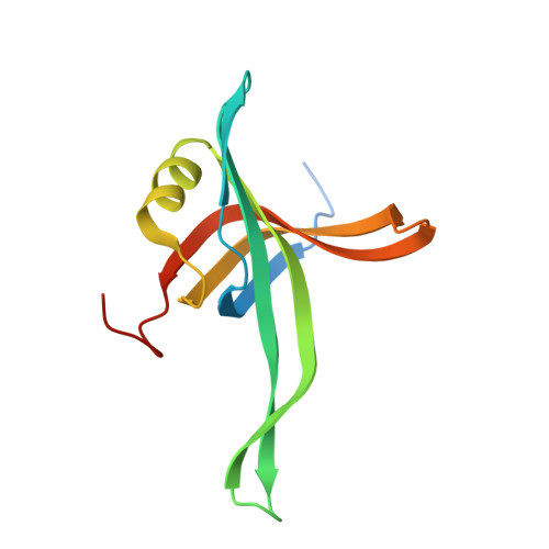
4DAM - PubMed Abstract:
The linear chromosome of Streptomyces coelicolor contains two paralogous ssb genes, ssbA and ssbB. Following mutational analysis, we concluded that ssbA is essential, whereas ssbB plays a key role in chromosome segregation during sporulation. In the ssbB mutant, ∼30% of spores lacked DNA. The two ssb genes were expressed differently; in minimal medium, gene expression was prolonged for both genes and significantly upregulated for ssbB. The ssbA gene is transcribed as part of a polycistronic mRNA from two initiation sites, 163 bp and 75 bp upstream of the rpsF translational start codon. The ssbB gene is transcribed as a monocistronic mRNA, from an unusual promoter region, 73 bp upstream of the AUG codon. Distinctive DNA-binding affinities of single-stranded DNA-binding proteins monitored by tryptophan fluorescent quenching and electrophoretic mobility shift were observed. The crystal structure of SsbB at 1.7 Å resolution revealed a common OB-fold, lack of the clamp-like structure conserved in SsbA and previously unpublished S-S bridges between the A/B and C/D subunits. This is the first report of the determination of paralogous single-stranded DNA-binding protein structures from the same organism. Phylogenetic analysis revealed frequent duplication of ssb genes in Actinobacteria, whereas their strong retention suggests that they are involved in important cellular functions.
- Division of Molecular Biology, Rudjer Boskovic Institute, Zagreb 10002, Croatia.
Organizational Affiliation: