4D9S | pdb_00004d9s

Crystal structure of Arabidopsis thaliana UVR8 (UV Resistance locus 8)


Experimental Data Snapshot

  • Method: X-RAY DIFFRACTION
  • Resolution: 1.70 Å
  • R-Value Free: 
    0.194 (Depositor), 0.186 (DCC) 
  • R-Value Work: 
    0.160 (Depositor), 0.155 (DCC) 
  • R-Value Observed: 
    0.161 (Depositor) 

Starting Model: experimental
View more details

wwPDB Validation 3D Report Full Report

Validation slider image for 4D9S

This is version 1.1 of the entry. See complete history

Literature

Plant UVR8 Photoreceptor Senses UV-B by Tryptophan-Mediated Disruption of Cross-Dimer Salt Bridges.

Christie, J.M.Arvai, A.S.Baxter, K.J.Heilmann, M.Pratt, A.J.O'Hara, A.Kelly, S.M.Hothorn, M.Smith, B.O.Hitomi, K.Jenkins, G.I.Getzoff, E.D.

(2012) Science 335: 1492-1496

  • DOI: https://doi.org/10.1126/science.1218091
  • Primary Citation Related Structures: 
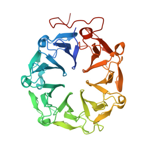
    4D9S

  • PubMed Abstract: 

    The recently identified plant photoreceptor UVR8 (UV RESISTANCE LOCUS 8) triggers regulatory changes in gene expression in response to ultraviolet-B (UV-B) light through an unknown mechanism. Here, crystallographic and solution structures of the UVR8 homodimer, together with mutagenesis and far-UV circular dichroism spectroscopy, reveal its mechanisms for UV-B perception and signal transduction. β-propeller subunits form a remarkable, tryptophan-dominated, dimer interface stitched together by a complex salt-bridge network. Salt-bridging arginines flank the excitonically coupled cross-dimer tryptophan "pyramid" responsible for UV-B sensing. Photoreception reversibly disrupts salt bridges, triggering dimer dissociation and signal initiation. Mutation of a single tryptophan to phenylalanine retunes the photoreceptor to detect UV-C wavelengths. Our analyses establish how UVR8 functions as a photoreceptor without a prosthetic chromophore to promote plant development and survival in sunlight.


  • Organizational Affiliation
    • Institute of Molecular, Cell and Systems Biology, College of Medical, Veterinary and Life Sciences, University of Glasgow, Glasgow, UK.

Macromolecule Content 

  • Total Structure Weight: 87.1 kDa 
  • Atom Count: 6,661 
  • Modeled Residue Count: 763 
  • Deposited Residue Count: 812 
  • Unique protein chains: 1

Macromolecules

Find similar proteins by:|  3D Structure
Entity ID: 1
MoleculeChains  Sequence LengthOrganismDetailsImage
UVB-resistance protein UVR8
A, B
406Arabidopsis thalianaMutation(s): 0 
Gene Names: UVR8At5g63860
UniProt
Find proteins for Q9FN03 (Arabidopsis thaliana)
Explore Q9FN03 
Go to UniProtKB:  Q9FN03
Entity Groups
Sequence Clusters30% Identity50% Identity70% Identity90% Identity95% Identity100% Identity
UniProt GroupQ9FN03
Sequence Annotations
Expand
Reference Sequence

Experimental Data & Validation

Experimental Data

  • Method: X-RAY DIFFRACTION
  • Resolution: 1.70 Å
  • R-Value Free:  0.194 (Depositor), 0.186 (DCC) 
  • R-Value Work:  0.160 (Depositor), 0.155 (DCC) 
  • R-Value Observed: 0.161 (Depositor) 
Space Group: P 1 21 1
Unit Cell:
Length ( Å )Angle ( ˚ )
a = 68.117α = 90
b = 51.511β = 102.89
c = 97.084γ = 90
Software Package:
Software NamePurpose
Blu-Icedata collection
PHASERphasing
PHENIXrefinement
HKL-2000data reduction
HKL-2000data scaling

Structure Validation

View Full Validation Report


Entry History 

Deposition Data

Revision History  (Full details and data files)

  • Version 1.0: 2012-04-04
    Type: Initial release
  • Version 1.1: 2023-09-13
    Changes: Data collection, Database references, Refinement description