4D47

X-ray structure of the levansucrase from Erwinia amylovora


Experimental Data Snapshot

  • Method: X-RAY DIFFRACTION
  • Resolution: 2.77 Å
  • R-Value Free: 0.298 
  • R-Value Work: 0.262 
  • R-Value Observed: 0.264 

wwPDB Validation   3D Report Full Report


Ligand Structure Quality Assessment 


This is version 1.4 of the entry. See complete history


Literature

The Crystal Structure of Erwinia Amylovora Levansucrase Provides a Snapshot of the Products of Sucrose Hydrolysis Trapped Into the Active Site.

Wuerges, J.Caputi, L.Cianci, M.Boivin, S.Meijers, R.Benini, S.

(2015) J Struct Biol 191: 290

  • DOI: https://doi.org/10.1016/j.jsb.2015.07.010
  • Primary Citation of Related Structures:  
    4D47

  • PubMed Abstract: 

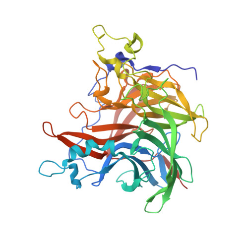
    Levansucrases are members of the glycoside hydrolase family and catalyse both the hydrolysis of the substrate sucrose and the transfer of fructosyl units to acceptor molecules. In the presence of sufficient sucrose, this may either lead to the production of fructooligosaccharides or fructose polymers. Aim of this study is to rationalise the differences in the polymerisation properties of bacterial levansucrases and in particular to identify structural features that determine different product spectrum in the levansucrase of the Gram-negative bacterium Erwinia amylovora (Ea Lsc, EC 2.4.1.10) as compared to Gram-positive bacteria such as Bacillus subtilis levansucrase. Ea is an enterobacterial pathogen responsible for the Fire Blight disease in rosaceous plants (e.g., apple and pear) with considerable interest for the agricultural industry. The crystal structure of Ea Lsc was solved at 2.77 Å resolution and compared to those of other fructosyltransferases from Gram-positive and Gram-negative bacteria. We propose the structural features, determining the different reaction products, to reside in just a few loops at the rim of the active site funnel. Moreover we propose that loop 8 may have a role in product length determination in Gluconacetobacter diazotrophicus LsdA and Microbacterium saccharophilum FFase. The Ea Lsc structure shows for the first time the products of sucrose hydrolysis still bound in the active site.


  • Organizational Affiliation

    Bioorganic Chemistry and Bio-Crystallography laboratory (B(2)Cl), Faculty of Science and Technology, Free University of Bolzano, 39100 Bolzano, Italy. Electronic address: jochen.wuerges@unibz.it.


Macromolecules
Find similar proteins by:  (by identity cutoff)  |  3D Structure
Entity ID: 1
MoleculeChains Sequence LengthOrganismDetailsImage
LEVANSUCRASE
A, B, C, D, E
A, B, C, D, E, F, G, H
415Erwinia amylovora ATCC 49946Mutation(s): 0 
EC: 2.4.1.10
UniProt
Find proteins for A0A0M3KKU6 (Erwinia amylovora (strain ATCC 49946 / CCPPB 0273 / Ea273 / 27-3))
Explore A0A0M3KKU6 
Go to UniProtKB:  A0A0M3KKU6
Entity Groups  
Sequence Clusters30% Identity50% Identity70% Identity90% Identity95% Identity100% Identity
UniProt GroupA0A0M3KKU6
Sequence Annotations
Expand
  • Reference Sequence
Small Molecules
Ligands 2 Unique
IDChains Name / Formula / InChI Key2D Diagram3D Interactions
GLC
Query on GLC

Download Ideal Coordinates CCD File 
J [auth A]
L [auth B]
N [auth C]
P [auth D]
R [auth E]
J [auth A],
L [auth B],
N [auth C],
P [auth D],
R [auth E],
T [auth F],
V [auth G],
X [auth H]
alpha-D-glucopyranose
C6 H12 O6
WQZGKKKJIJFFOK-DVKNGEFBSA-N
FRU
Query on FRU

Download Ideal Coordinates CCD File 
I [auth A]
K [auth B]
M [auth C]
O [auth D]
Q [auth E]
I [auth A],
K [auth B],
M [auth C],
O [auth D],
Q [auth E],
S [auth F],
U [auth G],
W [auth H]
beta-D-fructofuranose
C6 H12 O6
RFSUNEUAIZKAJO-ARQDHWQXSA-N
Experimental Data & Validation

Experimental Data

  • Method: X-RAY DIFFRACTION
  • Resolution: 2.77 Å
  • R-Value Free: 0.298 
  • R-Value Work: 0.262 
  • R-Value Observed: 0.264 
  • Space Group: P 21 21 2
Unit Cell:
Length ( Å )Angle ( ˚ )
a = 155.589α = 90
b = 178.736β = 90
c = 158.806γ = 90
Software Package:
Software NamePurpose
PHENIXrefinement
XDSdata reduction
SCALAdata scaling
BALBESphasing

Structure Validation

View Full Validation Report


Ligand Structure Quality Assessment 


Entry History 

Deposition Data

Revision History  (Full details and data files)

  • Version 1.0: 2015-08-05
    Type: Initial release
  • Version 1.1: 2015-09-09
    Changes: Database references
  • Version 1.2: 2018-03-07
    Changes: Data collection, Source and taxonomy
  • Version 1.3: 2020-07-29
    Type: Remediation
    Reason: Carbohydrate remediation
    Changes: Data collection, Derived calculations, Other, Structure summary
  • Version 1.4: 2023-12-20
    Changes: Data collection, Database references, Refinement description, Structure summary