4CR7

Crystal structure of the N-acetyl-D-mannosamine dehydrogenase with n-acetylmannosamine


Experimental Data Snapshot

  • Method: X-RAY DIFFRACTION
  • Resolution: 2.15 Å
  • R-Value Free: 0.231 
  • R-Value Work: 0.201 
  • R-Value Observed: 0.202 

wwPDB Validation   3D Report Full Report


Ligand Structure Quality Assessment 


This is version 1.3 of the entry. See complete history


Literature

Crystal Structures and Functional Studies Clarify Substrate Selectivity and Catalytic Residues for the Unique Orphan Enzyme N-Acetyl-D-Mannosamine Dehydrogenase.

Sola-Carvajal, A.Gil-Ortiz, F.Garcia-Carmona, F.Rubio, V.Sanchez-Ferrer, A.

(2014) Biochem J 462: 499

  • DOI: https://doi.org/10.1042/BJ20140266
  • Primary Citation of Related Structures:  
    4CR6, 4CR7, 4CR8

  • PubMed Abstract: 

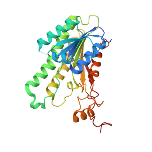
    NAMDH (N-acetyl-D-mannosamine dehydrogenase), from the soil bacteroidete Flavobacterium sp. 141-8, catalyses a rare NAD+-dependent oxidation of ManNAc (N-acetyl-D-mannosamine) into N-acetylmannosamino-lactone, which spontaneously hydrolyses into N-acetylmannosaminic acid. NAMDH belongs to the SDR (short-chain dehydrogenase/reductase) superfamily and is the only NAMDH characterized to date. Thorough functional, stability, site-directed mutagenesis and crystallographic studies have been carried out to understand better the structural and biochemical aspects of this unique enzyme. NAMDH exhibited a remarkable alkaline pH optimum (pH 9.4) with a high thermal stability in glycine buffer (Tm=64°C) and a strict selectivity towards ManNAc and NAD+. Crystal structures of ligand-free and ManNAc- and NAD+-bound enzyme forms revealed a compact homotetramer having point 222 symmetry, formed by subunits presenting the characteristic SDR α3β7α3 sandwich fold. A highly developed C-terminal tail used as a latch connecting nearby subunits stabilizes the tetramer. A dense network of polar interactions with the substrate including the encasement of its acetamido group in a specific binding pocket and the hydrogen binding of the sugar 4OH atom ensure specificity for ManNAc. The NAMDH-substrate complexes and site-directed mutagenesis studies identify the catalytic tetrad and provide useful traits for identifying new NAMDH sequences.


  • Organizational Affiliation

    *Department of Biochemistry and Molecular Biology-A, Faculty of Biology, Regional Campus of International Excellence "Campus Mare Nostrum", University of Murcia, Campus Espinardo, E-30100 Murcia, Spain.


Macromolecules
Find similar proteins by:  (by identity cutoff)  |  3D Structure
Entity ID: 1
MoleculeChains Sequence LengthOrganismDetailsImage
N-ACYLMANNOSAMINE 1-DEHYDROGENASE
A, B, C, D, E
A, B, C, D, E, F, G, H, I, J, K, L, M, N, O, P
271Flavobacterium sp. 141-8Mutation(s): 0 
EC: 1.1.1.233
UniProt
Find proteins for P22441 (Flavobacterium sp. (strain 141-8))
Explore P22441 
Go to UniProtKB:  P22441
Entity Groups  
Sequence Clusters30% Identity50% Identity70% Identity90% Identity95% Identity100% Identity
UniProt GroupP22441
Sequence Annotations
Expand
  • Reference Sequence
Small Molecules
Ligands 3 Unique
IDChains Name / Formula / InChI Key2D Diagram3D Interactions
BM3
Query on BM3

Download Ideal Coordinates CCD File 
CA [auth F]
GA [auth G]
IA [auth H]
KA [auth I]
MA [auth J]
CA [auth F],
GA [auth G],
IA [auth H],
KA [auth I],
MA [auth J],
OA [auth K],
PA [auth L],
QA [auth M],
R [auth A],
SA [auth N],
T [auth B],
U [auth C],
UA [auth O],
V [auth D],
WA [auth P],
Y [auth E]
2-acetamido-2-deoxy-alpha-D-mannopyranose
C8 H15 N O6
OVRNDRQMDRJTHS-UOLFYFMNSA-N
PG4
Query on PG4

Download Ideal Coordinates CCD File 
AA [auth F]
BA [auth F]
DA [auth F]
EA [auth F]
FA [auth G]
AA [auth F],
BA [auth F],
DA [auth F],
EA [auth F],
FA [auth G],
HA [auth H],
JA [auth I],
LA [auth J],
NA [auth K],
Q [auth A],
S [auth B],
VA [auth P],
X [auth E],
Z [auth F]
TETRAETHYLENE GLYCOL
C8 H18 O5
UWHCKJMYHZGTIT-UHFFFAOYSA-N
MAN
Query on MAN

Download Ideal Coordinates CCD File 
RA [auth M],
TA [auth N],
W [auth D],
XA [auth P]
alpha-D-mannopyranose
C6 H12 O6
WQZGKKKJIJFFOK-PQMKYFCFSA-N
Experimental Data & Validation

Experimental Data

  • Method: X-RAY DIFFRACTION
  • Resolution: 2.15 Å
  • R-Value Free: 0.231 
  • R-Value Work: 0.201 
  • R-Value Observed: 0.202 
  • Space Group: P 1
Unit Cell:
Length ( Å )Angle ( ˚ )
a = 99.39α = 67.43
b = 100.12β = 89.75
c = 111.6γ = 72.46
Software Package:
Software NamePurpose
REFMACrefinement
MOSFLMdata reduction
SCALAdata scaling
MOLREPphasing

Structure Validation

View Full Validation Report


Ligand Structure Quality Assessment 


Entry History 

Deposition Data

Revision History  (Full details and data files)

  • Version 1.0: 2014-07-09
    Type: Initial release
  • Version 1.1: 2014-09-03
    Changes: Atomic model, Database references
  • Version 1.2: 2020-07-29
    Type: Remediation
    Reason: Carbohydrate remediation
    Changes: Advisory, Data collection, Derived calculations, Other, Structure summary
  • Version 1.3: 2023-12-20
    Changes: Data collection, Database references, Refinement description, Structure summary