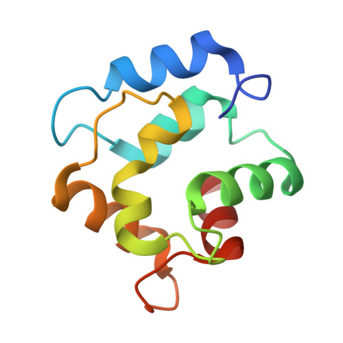
Refined crystal structure of calcium-liganded carp parvalbumin 4.25 at 1.5-A resolution.
Kumar, V.D., Lee, L., Edwards, B.F.(1990) Biochemistry 29: 1404-1412
- PubMed: 2334704 Search on PubMed
- DOI: https://doi.org/10.1021/bi00458a010
- Primary Citation Related Structures:
4CPV - PubMed Abstract:
The crystal structure of carp parvalbumin (pI = 4.25) has been refined by restrained least-squares analysis employing X-ray diffractometer data to 1.5-A resolution. The final residual for 12,653 reflections between 10 and 1.5 A with I(hkl) greater than 2 sigma(I) is 0.215. A total of 74 solvent molecules were included in the least-squares analysis. The root mean square deviation from ideality of bond lengths is 0.024 A. The model has a root mean square difference of 0.59 A from the positions of the main-chain atoms in a previously reported structure [Moews, P. C., & Kretsinger, R. H. (1975) J. Mol. Biol. 91, 201-228], which was refined by difference Fourier syntheses using data collected by film to 1.9 A. Although the overall features of the two models are very similar, there are significant differences in the amino-terminal region, which was extensively refit, and in the number of oxygen atoms liganding calcium in the CD and EF sites, which increased from six to seven in the CD site and decreased from eight to seven in the EF site.
- Department of Biochemistry, Wayne State University School of Medicine, Detroit, Michigan 48201.
Organizational Affiliation: