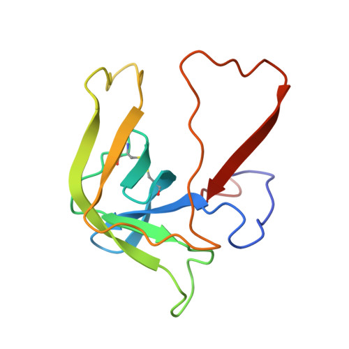
Structure of alpha-chymotrypsin refined at 1.68 A resolution.
Tsukada, H., Blow, D.M.(1985) J Mol Biol 184: 703-711
- PubMed: 4046030 Search on PubMed
- DOI: 10.1016/0022-2836(85)90314-6
- Primary Citation of Related Structures:  
4CHA - PubMed Abstract: 
- Structure and Mechanism of Chymotrypsin
Blow, D.M.
(1976) Acc Chem Res 9: 145 - The Active Centers of Serine Proteinases
Hartley, B.S.
(1974) Ann N Y Acad Sci 227: 438 - Comparison of the Crystal Structures of Chymotrypsinogen-A and Alpha-Chymotrypsin
Wright, H.T.
(1973) J Mol Biol 79: 1 - Structure of Crystalline Alpha-Chymotrypsin. V. The Atomic Structure of Tosyl-Alpha-Chymotrypsin at 2 Angstroms Resolution
Birktoft, J.J., Blow, D.M.
(1972) J Mol Biol 68: 187 - Structure of Crystalline Methyl-Chymotrypsin
Wright, C.S., Hess, G.P., Blow, D.M.
(1972) J Mol Biol 63: 295 - Alpha-Chymotrypsin,What Can We Learn About Catalysis from X-Ray Diffraction (Query).
Henderson, R., Wright, C.S., Hess, G.P., Blow, D.M.
(1972) Cold Spring Harb Symp Quant Biol 36: 63 - Chymotrypsin-Chemical Properties and Catalysis
Hess, G.P.
(1971) The Enzymes,third Edition 3: 213 - The Structure of Chymotrypsin
Blow, D.M.
(1971) The Enzymes,third Edition 3: 185 - Structure of Crystalline Alpha-Chymotrypsin. Iv. The Structure of Indoleacryloyl-Alpha-Chymotrypsin and its Relevance to the Hydrolytic Mechanism of the Enzyme
Henderson, R.
(1970) J Mol Biol 54: 341 - The Structure of Alpha-Chymotrypsin
Birktoft, J.J., Blow, D.M., Henderson, R., Steitz, T.A.
(1970) Philos Trans R Soc London,ser B 257: 67 - The Study of Alpha-Chymotrypsin by X-Ray Diffraction
Blow, D.M.
(1969) Biochem J 112: 261 - Structure of Crystalline Alpha-Chymotrypsin. III. Crystallographic Studies of Substrates and Inhibitors Bound to the Active Site of Alpha-Chymotrypsin
Steitz, T.A., Henderson, R., Blow, D.M.
(1969) J Mol Biol 46: 337 - Structure of Crystalline Alpha-Chymotrypsin. II. A Preliminary Report Including a Hypothesis for the Activation Mechanism
Sigler, P.B., Blow, D.M., Matthews, B.W., Henderson, R.
(1968) J Mol Biol 35: 143 - Three-Dimensional Structure of Tosyl-Alpha-Chymotrypsin
Matthews, B.W., Sigler, P.B., Henderson, R., Blow, D.M.
(1967) Nature 214: 652
Diffraction data for alpha-chymotrypsin crystals at -10 degrees C were measured at 1.68 A resolution and refined by restrained structure-factor least-squares refinement. The two independent chymotrypsin molecules in the crystallographic asymmetric unit were refined independently ...
Diffraction data for alpha-chymotrypsin crystals at -10 degrees C were measured at 1.68 A resolution and refined by restrained structure-factor least-squares refinement. The two independent chymotrypsin molecules in the crystallographic asymmetric unit were refined independently. The overall structure of alpha-chymotrypsin is little changed from published co-ordinates. The root-mean-square shift of C alpha co-ordinates is 0.42 A, co-ordinates for the two molecules showing a root-mean-square difference of 0.19 A. Certain regions with high disorder (residues 9 to 14, 73 to 79) remain difficult to interpret and several side-chains are disordered. Some water molecule positions have been changed. The absence of the tosyl group has made a significant difference to the refined structure at the active site. This now agrees closely with other enzymes of the trypsin family that have been refined at high resolution. There is a strong hydrogen bond between N epsilon 2 (His57) and O gamma (Ser195) in the free enzyme, in line with the published description of the charge relay system.
Related Citations: 
Organizational Affiliation: 
Department of Medicine, University of Alabama at Birmingham, Birmingham, AL 35294; paulg@uab.edu.