4BW7

Calmodulin in complex with strontium


Experimental Data Snapshot

  • Method: X-RAY DIFFRACTION
  • Resolution: 1.81 Å
  • R-Value Free: 0.251 
  • R-Value Work: 0.205 
  • R-Value Observed: 0.208 

wwPDB Validation   3D Report Full Report


This is version 2.1 of the entry. See complete history


Literature

Crystallographic Snapshots of Initial Steps in the Collapse of the Calmodulin Central Helix

Kursula, P.

(2014) Acta Crystallogr D Biol Crystallogr 70: 24

  • DOI: https://doi.org/10.1107/S1399004713024437
  • Primary Citation of Related Structures:  
    4BW7, 4BW8

  • PubMed Abstract: 

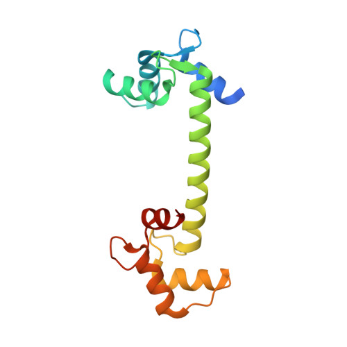
    Calmodulin is one of the most well characterized proteins and a widely used model system for calcium binding and large-scale protein conformational changes. Its long central helix is usually cut in half when a target peptide is bound. Here, two new crystal structures of calmodulin are presented, in which conformations possibly representing the first steps of calmodulin conformational collapse have been trapped. The central helix in the two structures is bent in the middle, causing a significant movement of the N- and C-terminal lobes with respect to one another. In both of the bent structures, a nearby polar side chain is inserted into the helical groove, disrupting backbone hydrogen bonding. The structures give an insight into the details of the factors that may be involved in the distortion of the central helix upon ligand peptide binding.


  • Organizational Affiliation

    Department of Biochemistry and Biocenter Oulu, University of Oulu, Oulu, Finland.


Macromolecules
Find similar proteins by:  (by identity cutoff)  |  3D Structure
Entity ID: 1
MoleculeChains Sequence LengthOrganismDetailsImage
CALMODULINA,
B [auth C]
149Homo sapiensMutation(s): 0 
UniProt & NIH Common Fund Data Resources
Find proteins for P0DP23 (Homo sapiens)
Explore P0DP23 
Go to UniProtKB:  P0DP23
PHAROS:  P0DP23
GTEx:  ENSG00000198668 
Entity Groups  
Sequence Clusters30% Identity50% Identity70% Identity90% Identity95% Identity100% Identity
UniProt GroupP0DP23
Sequence Annotations
Expand
  • Reference Sequence
Experimental Data & Validation

Experimental Data

  • Method: X-RAY DIFFRACTION
  • Resolution: 1.81 Å
  • R-Value Free: 0.251 
  • R-Value Work: 0.205 
  • R-Value Observed: 0.208 
  • Space Group: P 1
Unit Cell:
Length ( Å )Angle ( ˚ )
a = 24.34α = 88.51
b = 53.94β = 96.61
c = 59.37γ = 93.96
Software Package:
Software NamePurpose
PHENIXrefinement
XDSdata reduction
XSCALEdata scaling
PHASERphasing

Structure Validation

View Full Validation Report


Entry History 

Deposition Data

  • Released Date: 2014-01-15 
  • Deposition Author(s): Kursula, P.

Revision History  (Full details and data files)

  • Version 1.0: 2014-01-15
    Type: Initial release
  • Version 1.1: 2014-01-22
    Changes: Database references
  • Version 2.0: 2018-01-17
    Changes: Atomic model, Data collection
  • Version 2.1: 2023-12-20
    Changes: Data collection, Database references, Derived calculations, Other, Refinement description