4BB3

Isopenicillin N synthase with the dipeptide substrate analogue AhC


Experimental Data Snapshot

  • Method: X-RAY DIFFRACTION
  • Resolution: 1.40 Å
  • R-Value Free: 0.188 
  • R-Value Work: 0.172 
  • R-Value Observed: 0.173 

wwPDB Validation   3D Report Full Report


Ligand Structure Quality Assessment 


This is version 1.2 of the entry. See complete history


Literature

The Crystal Structure of Isopenicillin N Synthase with a Dipeptide Substrate Analogue.

Daruzzaman, A.Clifton, I.J.Adlington, R.M.Baldwin, J.E.Rutledge, P.J.

(2013) Arch Biochem Biophys 530: 48

  • DOI: https://doi.org/10.1016/j.abb.2012.12.012
  • Primary Citation of Related Structures:  
    4BB3

  • PubMed Abstract: 

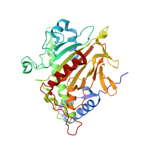
    Isopenicillin N synthase (IPNS) converts its linear tripeptide substrate δ-L-α-aminoadipoyl-L-cysteinyl-D-valine (ACV) to bicyclic isopenicillin N (IPN), the key step in penicillin biosynthesis. Solution-phase incubation experiments have shown that IPNS will accept and oxidise a diverse array of substrate analogues, including tripeptides that incorporate L-homocysteine as their second residue, and tripeptides with truncated side-chains at the third amino acid such as δ-L-α-aminoadipoyl-L-cysteinyl-D-α-aminobutyrate (ACAb), δ-L-α-aminoadipoyl-L-cysteinyl-D-alanine (ACA) and δ-L-α-aminoadipoyl-L-cysteinyl-glycine (ACG). However IPNS does not react with dipeptide substrates. To probe this selectivity we have crystallised the enzyme with the dipeptide δ-L-α-aminoadipoyl-L-homocysteine (AhC) and solved a crystal structure for the IPNS:Fe(II):AhC complex to 1.40 Å resolution. This structure reveals an unexpected mode of peptide binding at the IPNS active site, in which the homocysteinyl thiolate does not bind to iron. Instead the primary mode of binding sees the homocysteinyl carboxylate coordinated to the metal, while its side-chain is oriented into the region of the active site normally occupied by the benzyl group of protein residue Phe211.


  • Organizational Affiliation

    Chemistry Research Laboratory, University of Oxford, Mansfield Rd, Oxford OX1 3TA, UK.


Macromolecules
Find similar proteins by:  (by identity cutoff)  |  3D Structure
Entity ID: 1
MoleculeChains Sequence LengthOrganismDetailsImage
ISOPENICILLIN N SYNTHASE331Aspergillus nidulans FGSC A4Mutation(s): 0 
EC: 1.21.3.1
UniProt
Find proteins for P05326 (Emericella nidulans (strain FGSC A4 / ATCC 38163 / CBS 112.46 / NRRL 194 / M139))
Explore P05326 
Go to UniProtKB:  P05326
Entity Groups  
Sequence Clusters30% Identity50% Identity70% Identity90% Identity95% Identity100% Identity
UniProt GroupP05326
Sequence Annotations
Expand
  • Reference Sequence
Small Molecules
Ligands 3 Unique
IDChains Name / Formula / InChI Key2D Diagram3D Interactions
KKA
Query on KKA

Download Ideal Coordinates CCD File 
C [auth A](2S)-2-azanyl-6-oxidanylidene-6-[[(2S)-1-oxidanyl-1-oxidanylidene-4-sulfanyl-butan-2-yl]amino]hexanoic acid
C10 H18 N2 O5 S
SDIJIFRHLPSENI-BQBZGAKWSA-N
SO4
Query on SO4

Download Ideal Coordinates CCD File 
D [auth A]SULFATE ION
O4 S
QAOWNCQODCNURD-UHFFFAOYSA-L
FE
Query on FE

Download Ideal Coordinates CCD File 
B [auth A]FE (III) ION
Fe
VTLYFUHAOXGGBS-UHFFFAOYSA-N
Experimental Data & Validation

Experimental Data

  • Method: X-RAY DIFFRACTION
  • Resolution: 1.40 Å
  • R-Value Free: 0.188 
  • R-Value Work: 0.172 
  • R-Value Observed: 0.173 
  • Space Group: P 21 21 21
Unit Cell:
Length ( Å )Angle ( ˚ )
a = 46.508α = 90
b = 71.202β = 90
c = 100.884γ = 90
Software Package:
Software NamePurpose
REFMACrefinement
MOSFLMdata reduction
SCALAdata scaling

Structure Validation

View Full Validation Report


Ligand Structure Quality Assessment 


Entry History 

Deposition Data

Revision History  (Full details and data files)

  • Version 1.0: 2013-04-17
    Type: Initial release
  • Version 1.1: 2017-07-05
    Changes: Refinement description
  • Version 1.2: 2023-12-20
    Changes: Data collection, Database references, Derived calculations, Other, Refinement description