Structural Characterization of S100A15 Reveals a Novel Zinc Coordination Site Among S100 Proteins and Altered Surface Chemistry with Functional Implications for Receptor Binding.
Murray, J.I., Tonkin, M.L., Whiting, A.L., Peng, F., Farnell, B., Cullen, J.T., Hof, F., Boulanger, M.J.(2012) BMC Struct Biol 12: 16
- PubMed: 22747601 Search on PubMedSearch on PubMed Central
- DOI: https://doi.org/10.1186/1472-6807-12-16
- Primary Citation Related Structures:
4AQI, 4AQJ - PubMed Abstract:
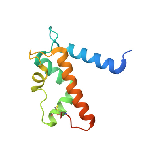
S100 proteins are a family of small, EF-hand containing calcium-binding signaling proteins that are implicated in many cancers. While the majority of human S100 proteins share 25-65% sequence similarity, S100A7 and its recently identified paralog, S100A15, display 93% sequence identity. Intriguingly, however, S100A7 and S100A15 serve distinct roles in inflammatory skin disease; S100A7 signals through the receptor for advanced glycation products (RAGE) in a zinc-dependent manner, while S100A15 signals through a yet unidentified G-protein coupled receptor in a zinc-independent manner. Of the seven divergent residues that differentiate S100A7 and S100A15, four cluster in a zinc-binding region and the remaining three localize to a predicted receptor-binding surface. To investigate the structural and functional consequences of these divergent clusters, we report the X-ray crystal structures of S100A15 and S100A7D24G, a hybrid variant where the zinc ligand Asp24 of S100A7 has been substituted with the glycine of S100A15, to 1.7 Å and 1.6 Å resolution, respectively. Remarkably, despite replacement of the Asp ligand, zinc binding is retained at the S100A15 dimer interface with distorted tetrahedral geometry and a chloride ion serving as an exogenous fourth ligand. Zinc binding was confirmed using anomalous difference maps and solution binding studies that revealed similar affinities of zinc for S100A15 and S100A7. Additionally, the predicted receptor-binding surface on S100A7 is substantially more basic in S100A15 without incurring structural rearrangement. Here we demonstrate that S100A15 retains the ability to coordinate zinc through incorporation of an exogenous ligand resulting in a unique zinc-binding site among S100 proteins. The altered surface chemistry between S100A7 and S100A15 that localizes to the predicted receptor binding site is likely responsible for the differential recognition of distinct protein targets. Collectively, these data provide novel insight into the structural and functional consequences of the divergent surfaces between S100A7 and S100A15 that may be exploited for targeted therapies.
- Department of Chemistry, University of Victoria, PO Box 3065STN CSC, Victoria, BC V8W 3P6, Canada.
Organizational Affiliation: