4AQ1

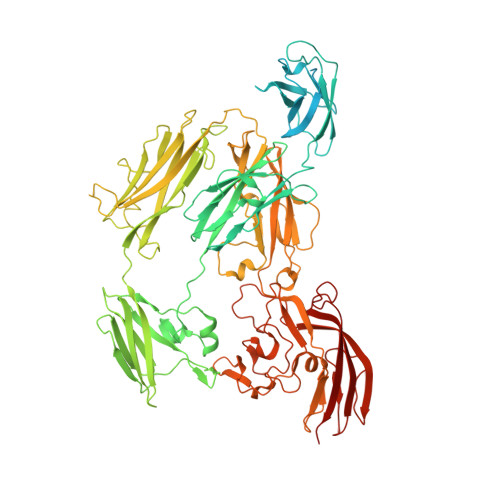
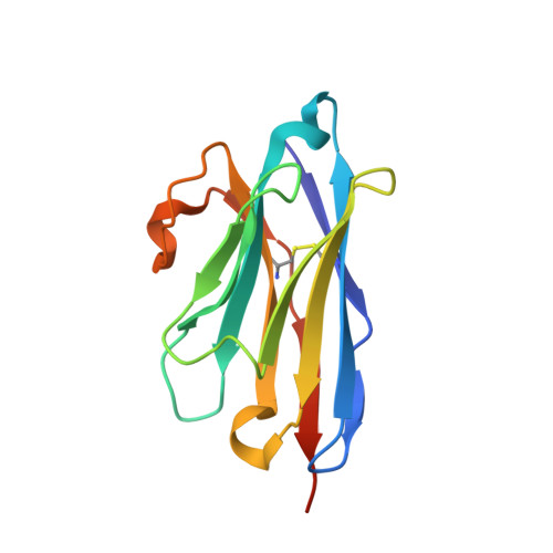
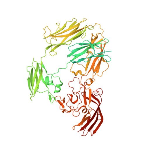
Structure of the SbsB S-layer protein of Geobacillus stearothermophilus PV72p2 in complex with nanobody KB6


Experimental Data Snapshot

  • Method: X-RAY DIFFRACTION
  • Resolution: 2.42 Å
  • R-Value Free: 0.236 
  • R-Value Work: 0.183 
  • R-Value Observed: 0.186 

wwPDB Validation   3D Report Full Report


This is version 1.2 of the entry. See complete history


Literature

Sbsb Structure and Lattice Reconstruction Unveil Ca21 Triggered S-Layer Assembly

Baranova, E.Fronzes, R.Garcia-Pino, A.Van Gerven, N.Papapostolou, D.Pehau-Arnaudet, G.Pardon, E.Steyaert, J.Howorka, S.Remaut, H.

(2012) Nature 487: 119

  • DOI: https://doi.org/10.1038/nature11155
  • Primary Citation of Related Structures:  
    4AQ1

  • PubMed Abstract: 

    S-layers are regular two-dimensional semipermeable protein layers that constitute a major cell-wall component in archaea and many bacteria. The nanoscale repeat structure of the S-layer lattices and their self-assembly from S-layer proteins (SLPs) have sparked interest in their use as patterning and display scaffolds for a range of nano-biotechnological applications. Despite their biological abundance and the technological interest in them, structural information about SLPs is limited to truncated and assembly-negative proteins. Here we report the X-ray structure of the SbsB SLP of Geobacillus stearothermophilus PV72/p2 by the use of nanobody-aided crystallization. SbsB consists of a seven-domain protein, formed by an amino-terminal cell-wall attachment domain and six consecutive immunoglobulin-like domains, that organize into a φ-shaped disk-like monomeric crystallization unit stabilized by interdomain Ca(2+) ion coordination. A Ca(2+)-dependent switch to the condensed SbsB quaternary structure pre-positions intermolecular contact zones and renders the protein competent for S-layer assembly. On the basis of crystal packing, chemical crosslinking data and cryo-electron microscopy projections, we present a model for the molecular organization of this SLP into a porous protein sheet inside the S-layer. The SbsB lattice represents a previously undescribed structural model for protein assemblies and may advance our understanding of SLP physiology and self-assembly, as well as the rational design of engineered higher-order structures for biotechnology.


  • Organizational Affiliation

    Structural and Molecular Microbiology, VIB Department of Structural Biology, VIB, Pleinlaan 2, 1050 Brussels, Belgium.


Macromolecules
Find similar proteins by:  (by identity cutoff)  |  3D Structure
Entity ID: 1
MoleculeChains Sequence LengthOrganismDetailsImage
SBSB PROTEIN892Geobacillus stearothermophilusMutation(s): 0 
UniProt
Find proteins for Q45664 (Geobacillus stearothermophilus)
Explore Q45664 
Go to UniProtKB:  Q45664
Entity Groups  
Sequence Clusters30% Identity50% Identity70% Identity90% Identity95% Identity100% Identity
UniProt GroupQ45664
Sequence Annotations
Expand
  • Reference Sequence
Find similar proteins by:  (by identity cutoff)  |  3D Structure
Entity ID: 2
MoleculeChains Sequence LengthOrganismDetailsImage
NBKB6
B, D
130Lama glamaMutation(s): 0 
Entity Groups  
Sequence Clusters30% Identity50% Identity70% Identity90% Identity95% Identity100% Identity
Sequence Annotations
Expand
  • Reference Sequence
Find similar proteins by:  (by identity cutoff)  |  3D Structure
Entity ID: 3
MoleculeChains Sequence LengthOrganismDetailsImage
SBSB PROTEIN892Geobacillus stearothermophilusMutation(s): 0 
UniProt
Find proteins for Q45664 (Geobacillus stearothermophilus)
Explore Q45664 
Go to UniProtKB:  Q45664
Entity Groups  
Sequence Clusters30% Identity50% Identity70% Identity90% Identity95% Identity100% Identity
UniProt GroupQ45664
Sequence Annotations
Expand
  • Reference Sequence
Experimental Data & Validation

Experimental Data

  • Method: X-RAY DIFFRACTION
  • Resolution: 2.42 Å
  • R-Value Free: 0.236 
  • R-Value Work: 0.183 
  • R-Value Observed: 0.186 
  • Space Group: P 1
Unit Cell:
Length ( Å )Angle ( ˚ )
a = 68.5α = 112.5
b = 88.5β = 101.6
c = 95.2γ = 84.7
Software Package:
Software NamePurpose
REFMACrefinement
XDSdata reduction
XSCALEdata scaling
PHENIXphasing

Structure Validation

View Full Validation Report


Entry History 

Deposition Data

Revision History  (Full details and data files)

  • Version 1.0: 2012-06-13
    Type: Initial release
  • Version 1.1: 2012-07-11
    Changes: Other
  • Version 1.2: 2012-10-03
    Changes: Derived calculations