4AQ1 | pdb_00004aq1

Structure of the SbsB S-layer protein of Geobacillus stearothermophilus PV72p2 in complex with nanobody KB6


Experimental Data Snapshot

  • Method: X-RAY DIFFRACTION
  • Resolution: 2.42 Å
  • R-Value Free: 
    0.236 (Depositor), 0.235 (DCC) 
  • R-Value Work: 
    0.183 (Depositor), 0.186 (DCC) 
  • R-Value Observed: 
    0.186 (Depositor) 

wwPDB Validation 3D Report Full Report

Validation slider image for 4AQ1

This is version 1.3 of the entry. See complete history

Literature

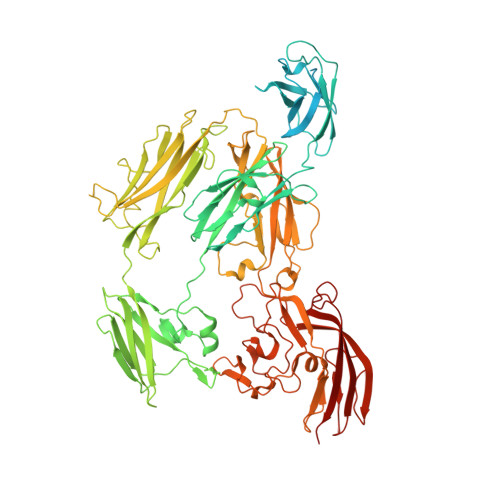
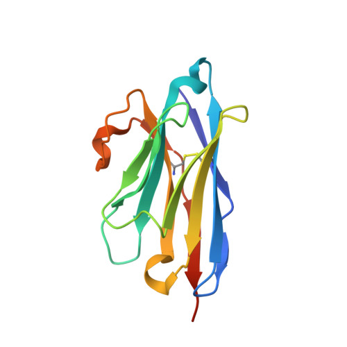
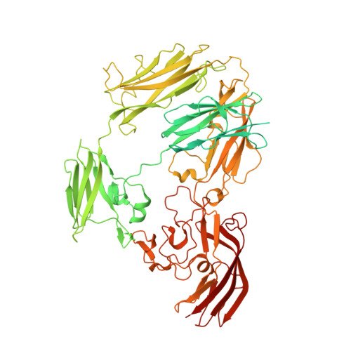
Sbsb Structure and Lattice Reconstruction Unveil Ca21 Triggered S-Layer Assembly

Baranova, E.Fronzes, R.Garcia-Pino, A.Van Gerven, N.Papapostolou, D.Pehau-Arnaudet, G.Pardon, E.Steyaert, J.Howorka, S.Remaut, H.

(2012) Nature 487: 119

  • DOI: https://doi.org/10.1038/nature11155
  • Primary Citation Related Structures: 
    4AQ1

  • PubMed Abstract: 

    S-layers are regular two-dimensional semipermeable protein layers that constitute a major cell-wall component in archaea and many bacteria. The nanoscale repeat structure of the S-layer lattices and their self-assembly from S-layer proteins (SLPs) have sparked interest in their use as patterning and display scaffolds for a range of nano-biotechnological applications. Despite their biological abundance and the technological interest in them, structural information about SLPs is limited to truncated and assembly-negative proteins. Here we report the X-ray structure of the SbsB SLP of Geobacillus stearothermophilus PV72/p2 by the use of nanobody-aided crystallization. SbsB consists of a seven-domain protein, formed by an amino-terminal cell-wall attachment domain and six consecutive immunoglobulin-like domains, that organize into a φ-shaped disk-like monomeric crystallization unit stabilized by interdomain Ca(2+) ion coordination. A Ca(2+)-dependent switch to the condensed SbsB quaternary structure pre-positions intermolecular contact zones and renders the protein competent for S-layer assembly. On the basis of crystal packing, chemical crosslinking data and cryo-electron microscopy projections, we present a model for the molecular organization of this SLP into a porous protein sheet inside the S-layer. The SbsB lattice represents a previously undescribed structural model for protein assemblies and may advance our understanding of SLP physiology and self-assembly, as well as the rational design of engineered higher-order structures for biotechnology.


  • Organizational Affiliation
    • Structural and Molecular Microbiology, VIB Department of Structural Biology, VIB, Pleinlaan 2, 1050 Brussels, Belgium.

Macromolecule Content 

  • Total Structure Weight: 218.24 kDa 
  • Atom Count: 12,335 
  • Modeled Residue Count: 1,596 
  • Deposited Residue Count: 2,044 
  • Unique protein chains: 3

Macromolecules

Find similar proteins by:|  3D Structure
Entity ID: 1
MoleculeChains  Sequence LengthOrganismDetailsImage
SBSB PROTEIN892Geobacillus stearothermophilusMutation(s): 0 
UniProt
Find proteins for Q45664 (Geobacillus stearothermophilus)
Explore Q45664 
Go to UniProtKB:  Q45664
Entity Groups
Sequence Clusters30% Identity50% Identity70% Identity90% Identity95% Identity100% Identity
UniProt GroupQ45664
Sequence Annotations
Expand
Reference Sequence
Find similar proteins by:|  3D Structure
Entity ID: 2
MoleculeChains  Sequence LengthOrganismDetailsImage
NBKB6
B, D
130Lama glamaMutation(s): 0 
Find similar proteins by:|  3D Structure
Entity ID: 3
MoleculeChains  Sequence LengthOrganismDetailsImage
SBSB PROTEIN892Geobacillus stearothermophilusMutation(s): 0 
UniProt
Find proteins for Q45664 (Geobacillus stearothermophilus)
Explore Q45664 
Go to UniProtKB:  Q45664
Entity Groups
Sequence Clusters30% Identity50% Identity70% Identity90% Identity95% Identity100% Identity
UniProt GroupQ45664
Sequence Annotations
Expand
Reference Sequence

Experimental Data & Validation

Experimental Data

  • Method: X-RAY DIFFRACTION
  • Resolution: 2.42 Å
  • R-Value Free:  0.236 (Depositor), 0.235 (DCC) 
  • R-Value Work:  0.183 (Depositor), 0.186 (DCC) 
  • R-Value Observed: 0.186 (Depositor) 
Space Group: P 1
Unit Cell:
Length ( Å )Angle ( ˚ )
a = 68.5α = 112.5
b = 88.5β = 101.6
c = 95.2γ = 84.7
Software Package:
Software NamePurpose
REFMACrefinement
XDSdata reduction
XSCALEdata scaling
PHENIXphasing

Structure Validation

View Full Validation Report


Entry History 

Deposition Data

Revision History  (Full details and data files)

  • Version 1.0: 2012-06-13
    Type: Initial release
  • Version 1.1: 2012-07-11
    Changes: Other
  • Version 1.2: 2012-10-03
    Changes: Derived calculations
  • Version 1.3: 2024-11-20
    Changes: Data collection, Database references, Derived calculations, Other, Refinement description, Structure summary