4AFY

Crystal structure of the FimX EAL domain in complex with reaction product pGpG


Experimental Data Snapshot

  • Method: X-RAY DIFFRACTION
  • Resolution: 2.01 Å
  • R-Value Free: 0.239 
  • R-Value Work: 0.208 
  • R-Value Observed: 0.210 

wwPDB Validation 3D Report Full Report


This is version 1.2 of the entry. See complete history


Literature

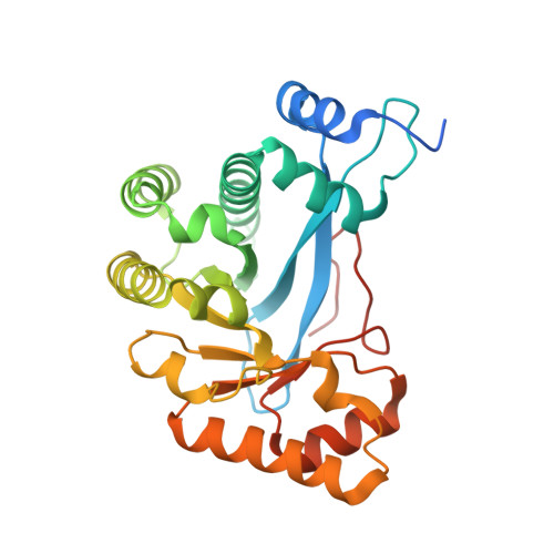
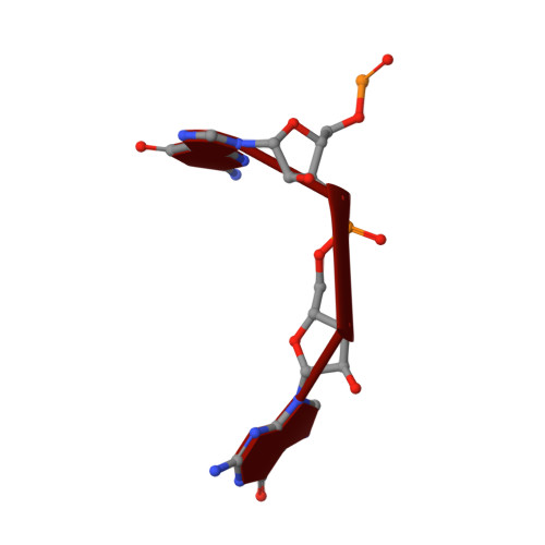
Crystal Structure of an Eal Domain in Complex with Reaction Product 5'-Pgpg

Robert-Paganin, J.Nonin-Lecomte, S.Rety, S.

(2012) PLoS One 7: 52424

  • DOI: 10.1371/journal.pone.0052424
  • Structures With Same Primary Citation

  • PubMed Abstract: 
  • FimX is a large multidomain protein containing an EAL domain and involved in twitching motility in Pseudomonas aeruginosa. We present here two crystallographic structures of the EAL domain of FimX (residues 438-686): one of the apo form and the other ...

    FimX is a large multidomain protein containing an EAL domain and involved in twitching motility in Pseudomonas aeruginosa. We present here two crystallographic structures of the EAL domain of FimX (residues 438-686): one of the apo form and the other of a complex with 5'-pGpG, the reaction product of the hydrolysis of c-di-GMP. In both crystal forms, the EAL domains form a dimer delimiting a large cavity encompassing the catalytic pockets. The ligand is trapped in this cavity by its sugar phosphate moiety. We confirmed by NMR that the guanine bases are not involved in the interaction in solution. We solved here the first structure of an EAL domain bound to the reaction product 5'-pGpG. Though isolated FimX EAL domain has a very low catalytic activity, which would not be significant compared to other catalytic EAL domains, the structure with the product of the reaction can provides some hints in the mechanism of hydrolysis of the c-di-GMP by EAL domains.


    Organizational Affiliation

    Laboratoire de Cristallographie et RMN biologiques, UMR 8015-Cente National de la Recherche Scientifique, Paris, France.


Macromolecules

Find similar proteins by: Sequence  |  Structure

Entity ID: 1
MoleculeChainsSequence LengthOrganismDetails
FIMX
A, B
274Pseudomonas aeruginosa PAO1Mutation(s): 0 
Gene Names: PA4959fimX
EC: 3.1.4.52
Find proteins for Q9HUK6 (Pseudomonas aeruginosa (strain ATCC 15692 / DSM 22644 / CIP 104116 / JCM 14847 / LMG 12228 / 1C / PRS 101 / PAO1))
Go to UniProtKB:  Q9HUK6
  • Find similar nucleic acids by: Sequence   |   Structure
Entity ID: 2
MoleculeChainsLengthOrganism
5'-R(*GP*GP)-3'C, D2synthetic construct
Small Molecules
Ligands 2 Unique
IDChainsName / Formula / InChI Key2D Diagram3D Interactions
MG
Query on MG

Download CCD File 
A, B
MAGNESIUM ION
Mg
JLVVSXFLKOJNIY-UHFFFAOYSA-N
 Ligand Interaction
NA
Query on NA

Download CCD File 
A
SODIUM ION
Na
FKNQFGJONOIPTF-UHFFFAOYSA-N
 Ligand Interaction
Experimental Data & Validation

Experimental Data

  • Method: X-RAY DIFFRACTION
  • Resolution: 2.01 Å
  • R-Value Free: 0.239 
  • R-Value Work: 0.208 
  • R-Value Observed: 0.210 
  • Space Group: P 43 21 2
Unit Cell:
Length ( Å )Angle ( ˚ )
a = 104.33α = 90
b = 104.33β = 90
c = 154.24γ = 90
Software Package:
Software NamePurpose
PHENIXrefinement
XDSdata reduction
XDSdata scaling
BALBESphasing

Structure Validation

View Full Validation Report


Entry History 

Deposition Data

Revision History 

  • Version 1.0: 2013-01-09
    Type: Initial release
  • Version 1.1: 2013-01-30
    Changes: Database references
  • Version 1.2: 2015-10-21
    Changes: Source and taxonomy