Biochemical and Structural Characterization of Two Dictyostelium Cellobiohydrolases from the Amoebozoa Kingdom Reveal a High Conservation between Distant Phylogenetic Trees of Life.
Hobdey, S.E., Knott, B.C., Haddad Momeni, M., Taylor, L.E., Borisova, A.S., Podkaminer, K.K., VanderWall, T.A., Himmel, M.E., Decker, S.R., Beckham, G.T., Stahlberg, J.(2016) Appl Environ Microbiol 82: 3395-4409
- PubMed: 27037126 Search on PubMedSearch on PubMed Central
- DOI: https://doi.org/10.1128/AEM.00163-16
- Primary Citation Related Structures:
4ZZP, 4ZZQ - PubMed Abstract:
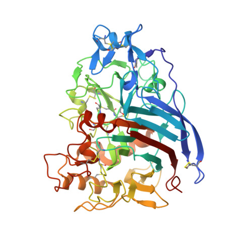
Glycoside hydrolase family 7 (GH7) cellobiohydrolases (CBHs) are enzymes commonly employed in plant cell wall degradation across eukaryotic kingdoms of life, as they provide significant hydrolytic potential in cellulose turnover. To date, many fungal GH7 CBHs have been examined, yet many questions regarding structure-activity relationships in these important natural and commercial enzymes remain. Here, we present the crystal structures and a biochemical analysis of two GH7 CBHs from social amoeba: Dictyostelium discoideum Cel7A (DdiCel7A) and Dictyostelium purpureum Cel7A (DpuCel7A). DdiCel7A and DpuCel7A natively consist of a catalytic domain and do not exhibit a carbohydrate-binding module (CBM). The structures of DdiCel7A and DpuCel7A, resolved to 2.1 Å and 2.7 Å, respectively, are homologous to those of other GH7 CBHs with an enclosed active-site tunnel. Two primary differences between the Dictyostelium CBHs and the archetypal model GH7 CBH, Trichoderma reesei Cel7A (TreCel7A), occur near the hydrolytic active site and the product-binding sites. To compare the activities of these enzymes with the activity of TreCel7A, the family 1 TreCel7A CBM and linker were added to the C terminus of each of the Dictyostelium enzymes, creating DdiCel7ACBM and DpuCel7ACBM, which were recombinantly expressed in T. reesei DdiCel7ACBM and DpuCel7ACBM hydrolyzed Avicel, pretreated corn stover, and phosphoric acid-swollen cellulose as efficiently as TreCel7A when hydrolysis was compared at their temperature optima. The Ki of cellobiose was significantly higher for DdiCel7ACBM and DpuCel7ACBM than for TreCel7A: 205, 130, and 29 μM, respectively. Taken together, the present study highlights the remarkable degree of conservation of the activity of these key natural and industrial enzymes across quite distant phylogenetic trees of life. GH7 CBHs are among the most important cellulolytic enzymes both in nature and for emerging industrial applications for cellulose breakdown. Understanding the diversity of these key industrial enzymes is critical to engineering them for higher levels of activity and greater stability. The present work demonstrates that two GH7 CBHs from social amoeba are surprisingly quite similar in structure and activity to the canonical GH7 CBH from the model biomass-degrading fungus T. reesei when tested under equivalent conditions (with added CBM-linker domains) on an industrially relevant substrate.
- Biosciences Center, National Renewable Energy Laboratory, Golden, Colorado, USA.
Organizational Affiliation: