4ZXW | pdb_00004zxw

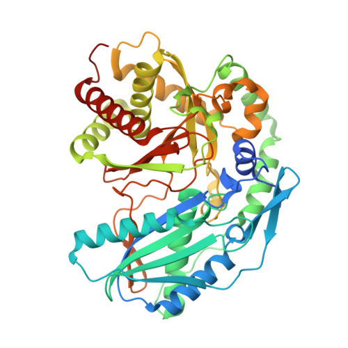
Crystal structure of SgcC5 protein from Streptomyces globisporus (complex with (R)-(-)-1-(2-naphthyl)-1,2-ethanediol and sucrose)


Experimental Data Snapshot

  • Method: X-RAY DIFFRACTION
  • Resolution: 2.19 Å
  • R-Value Free: 
    0.208 (Depositor), 0.211 (DCC) 
  • R-Value Work: 
    0.161 (Depositor), 0.170 (DCC) 
  • R-Value Observed: 
    0.162 (Depositor) 

Starting Model: experimental
View more details

wwPDB Validation 3D Report Full Report

Validation slider image for 4ZXW

Ligand Structure Quality Assessment 


This is version 2.1 of the entry. See complete history

Macromolecule Content 

  • Total Structure Weight: 103.33 kDa 
  • Atom Count: 7,291 
  • Modeled Residue Count: 886 
  • Deposited Residue Count: 924 
  • Unique protein chains: 1

Macromolecules

Find similar proteins by:|  3D Structure
Entity ID: 1
MoleculeChains  Sequence LengthOrganismDetailsImage
C-domain type II peptide synthetase
A, B
462Streptomyces globisporusMutation(s): 0 
UniProt
Find proteins for Q8GMG2 (Streptomyces globisporus)
Explore Q8GMG2 
Go to UniProtKB:  Q8GMG2
Entity Groups
Sequence Clusters30% Identity50% Identity70% Identity90% Identity95% Identity100% Identity
UniProt GroupQ8GMG2
Sequence Annotations
Expand
Reference Sequence

Oligosaccharides

Help  
Entity ID: 2
MoleculeChains Length2D Diagram GlycosylationD Interactions
beta-D-fructofuranose-(2-1)-alpha-D-glucopyranose
C, D
2N/A
Glycosylation Resources
GlyTouCan: G05551OP
GlyCosmos: G05551OP

Small Molecules

Ligands 3 Unique
IDChains Name / Formula / InChI Key2D Diagram3D Interactions
CXS

Query on CXS


Download:Ideal Coordinates CCD File
L [auth B]3-CYCLOHEXYL-1-PROPYLSULFONIC ACID
C9 H19 N O3 S
PJWWRFATQTVXHA-UHFFFAOYSA-N
4T7

Query on 4T7


Download:Ideal Coordinates CCD File
E [auth A],
I [auth B]
(1R)-1-(naphthalen-2-yl)ethane-1,2-diol
C12 H12 O2
QFNOPZNUHSDHKZ-LBPRGKRZSA-N
SO4

Query on SO4


Download:Ideal Coordinates CCD File
F [auth A],
G [auth A],
H [auth A],
J [auth B],
K [auth B]
SULFATE ION
O4 S
QAOWNCQODCNURD-UHFFFAOYSA-L

Biologically Interesting Molecules (External Reference) 

1 Unique

Experimental Data & Validation

Experimental Data

  • Method: X-RAY DIFFRACTION
  • Resolution: 2.19 Å
  • R-Value Free:  0.208 (Depositor), 0.211 (DCC) 
  • R-Value Work:  0.161 (Depositor), 0.170 (DCC) 
  • R-Value Observed: 0.162 (Depositor) 
Space Group: P 21 21 21
Unit Cell:
Length ( Å )Angle ( ˚ )
a = 99.397α = 90
b = 105.304β = 90
c = 108.167γ = 90
Software Package:
Software NamePurpose
PHENIXrefinement
HKL-3000data reduction
HKL-3000data scaling
PHASERphasing

Structure Validation

View Full Validation Report


Ligand Structure Quality Assessment 


Entry History 

& Funding Information

Deposition Data


Funding OrganizationLocationGrant Number
National Institutes of Health/National Institute of General Medical Sciences (NIH/NIGMS)United StatesGM094585
National Institutes of Health/National Institute of General Medical Sciences (NIH/NIGMS)United StatesGM098248

Revision History  (Full details and data files)

  • Version 1.0: 2015-06-17
    Type: Initial release
  • Version 1.1: 2016-09-21
    Changes: Source and taxonomy
  • Version 1.2: 2017-09-06
    Changes: Author supporting evidence, Derived calculations
  • Version 1.3: 2019-12-25
    Changes: Author supporting evidence
  • Version 2.0: 2020-07-29
    Type: Remediation
    Reason: Carbohydrate remediation
    Changes: Atomic model, Data collection, Derived calculations, Non-polymer description, Structure summary
  • Version 2.1: 2023-09-27
    Changes: Data collection, Database references, Refinement description, Structure summary