4ZQE

Crystal structure of DOX-P Reductoisomerase in complex with magnesium


Experimental Data Snapshot

  • Method: X-RAY DIFFRACTION
  • Resolution: 1.98 Å
  • R-Value Free: 0.195 
  • R-Value Work: 0.171 
  • R-Value Observed: 0.172 

wwPDB Validation   3D Report Full Report


This is version 1.0 of the entry. See complete history


Literature

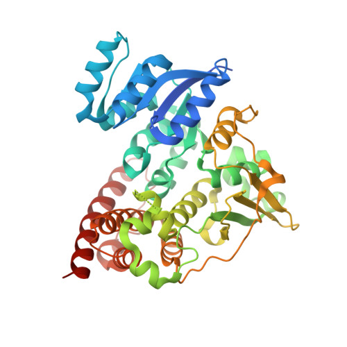
Crystal structures of the Moraxella catarrhalis DOX-P Reductoisomerase

Birkinshaw, R.W.Brady, R.L.

To be published.

Macromolecules
Find similar proteins by:  (by identity cutoff)  |  3D Structure
Entity ID: 1
MoleculeChains Sequence LengthOrganismDetailsImage
1-deoxy-D-xylulose 5-phosphate reductoisomerase
A, B
432Moraxella catarrhalisMutation(s): 0 
Gene Names: dxrDR90_1378
EC: 1.1.1.267
Entity Groups  
Sequence Clusters30% Identity50% Identity70% Identity90% Identity95% Identity100% Identity
Sequence Annotations
Expand
  • Reference Sequence
Small Molecules
Ligands 2 Unique
IDChains Name / Formula / InChI Key2D Diagram3D Interactions
SO4
Query on SO4

Download Ideal Coordinates CCD File 
C [auth A]
D [auth A]
E [auth A]
F [auth A]
I [auth B]
C [auth A],
D [auth A],
E [auth A],
F [auth A],
I [auth B],
J [auth B],
K [auth B],
L [auth B],
M [auth B]
SULFATE ION
O4 S
QAOWNCQODCNURD-UHFFFAOYSA-L
GOL
Query on GOL

Download Ideal Coordinates CCD File 
G [auth A],
H [auth A],
N [auth B],
O [auth B],
P [auth B]
GLYCEROL
C3 H8 O3
PEDCQBHIVMGVHV-UHFFFAOYSA-N
Experimental Data & Validation

Experimental Data

  • Method: X-RAY DIFFRACTION
  • Resolution: 1.98 Å
  • R-Value Free: 0.195 
  • R-Value Work: 0.171 
  • R-Value Observed: 0.172 
  • Space Group: P 41
Unit Cell:
Length ( Å )Angle ( ˚ )
a = 133.21α = 90
b = 133.21β = 90
c = 77.51γ = 90
Software Package:
Software NamePurpose
REFMACrefinement
XDSdata reduction
SCALAdata scaling
PHASERphasing

Structure Validation

View Full Validation Report


Entry History 

Deposition Data

Revision History  (Full details and data files)

  • Version 1.0: 2016-06-29
    Type: Initial release