4ZOR | pdb_00004zor

The structure of the S37P MS2 viral capsid assembly.


Experimental Data Snapshot

  • Method: X-RAY DIFFRACTION
  • Resolution: 2.20 Å
  • R-Value Free: 
    0.242 (Depositor), 0.243 (DCC) 
  • R-Value Work: 
    0.218 (Depositor), 0.221 (DCC) 
  • R-Value Observed: 
    0.219 (Depositor) 

Starting Model: experimental
View more details

wwPDB Validation 3D Report Full Report

Validation slider image for 4ZOR

This is version 1.2 of the entry. See complete history

Literature

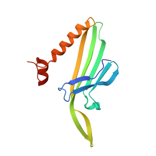
A Selection for Assembly Reveals That a Single Amino Acid Mutant of the Bacteriophage MS2 Coat Protein Forms a Smaller Virus-like Particle.

Asensio, M.A.Morella, N.M.Jakobson, C.M.Hartman, E.C.Glasgow, J.E.Sankaran, B.Zwart, P.H.Tullman-Ercek, D.

(2016) Nano Lett 16: 5944-5950

  • DOI: https://doi.org/10.1021/acs.nanolett.6b02948
  • Primary Citation Related Structures: 
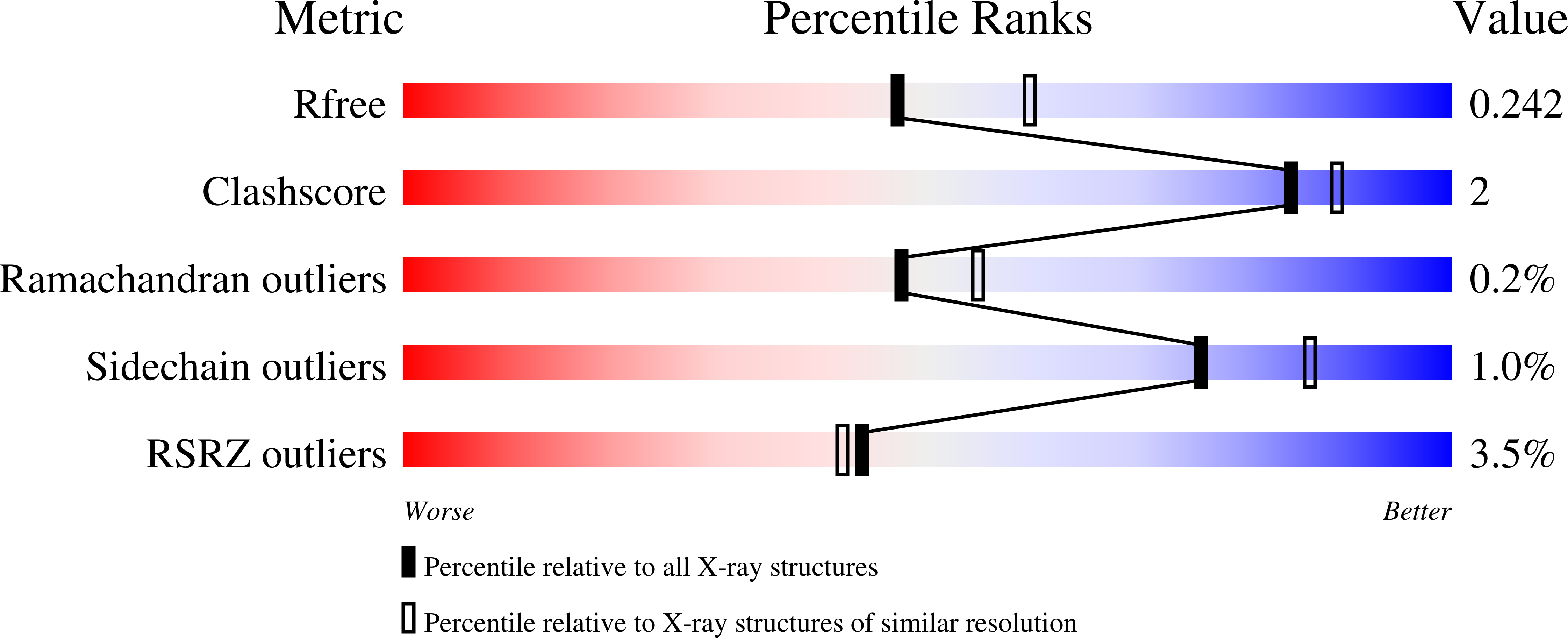
    4ZOR

  • PubMed Abstract: 

    Virus-like particles are used to encapsulate drugs, imaging agents, enzymes, and other biologically active molecules in order to enhance their function. However, the size of most virus-like particles is inflexible, precluding the design of appropriately sized containers for different applications. Here, we describe a chromatographic selection for virus-like particle assembly. Using this selection, we identified a single amino acid substitution to the coat protein of bacteriophage MS2 that mediates a uniform switch in particle geometry from T = 3 to T = 1 icosahedral symmetry. The resulting smaller particle retains the ability to be disassembled and reassembled in vitro and to be chemically modified to load cargo into its interior cavity. The pair of 27 and 17 nm MS2 particles will allow direct examination of the effect of size on function in established applications of virus-like particles, including drug delivery and imaging.


  • Organizational Affiliation
    • Department of Chemical and Biological Engineering, Northwestern University , Evanston, Illinois 60091, United States.

Macromolecule Content 

  • Total Structure Weight: 69.12 kDa 
  • Atom Count: 5,204 
  • Modeled Residue Count: 630 
  • Deposited Residue Count: 645 
  • Unique protein chains: 1

Macromolecules

Find similar proteins by:|  3D Structure
Entity ID: 1
MoleculeChains  Sequence LengthOrganismDetailsImage
Coat protein
A, B, C, D, E
129Emesvirus zinderiMutation(s): 1 
UniProt
Find proteins for P03612 (Escherichia phage MS2)
Explore P03612 
Go to UniProtKB:  P03612
Entity Groups
Sequence Clusters30% Identity50% Identity70% Identity90% Identity95% Identity100% Identity
UniProt GroupP03612
Sequence Annotations
Expand
Reference Sequence

Experimental Data & Validation

Experimental Data

  • Method: X-RAY DIFFRACTION
  • Resolution: 2.20 Å
  • R-Value Free:  0.242 (Depositor), 0.243 (DCC) 
  • R-Value Work:  0.218 (Depositor), 0.221 (DCC) 
  • R-Value Observed: 0.219 (Depositor) 
Space Group: F 2 3
Unit Cell:
Length ( Å )Angle ( ˚ )
a = 228.125α = 90
b = 228.125β = 90
c = 228.125γ = 90
Software Package:
Software NamePurpose
PHENIXrefinement
HKL-2000data reduction
HKL-2000data scaling
PHENIXphasing

Structure Validation

View Full Validation Report


Entry History 

Deposition Data

Revision History  (Full details and data files)

  • Version 1.0: 2016-09-07
    Type: Initial release
  • Version 1.1: 2016-09-28
    Changes: Database references
  • Version 1.2: 2023-09-27
    Changes: Data collection, Database references, Derived calculations, Refinement description