Functional and Structural Analysis of a beta-Glucosidase Involved in beta-1,2-Glucan Metabolism in Listeria innocua
Nakajima, M., Yoshida, R., Miyanaga, A., Abe, K., Takahashi, Y., Sugimoto, N., Toyoizumi, H., Nakai, H., Kitaoka, M., Taguchi, H.(2016) PLoS One 11: e0148870-e0148870
- PubMed: 26886583 Search on PubMedSearch on PubMed Central
- DOI: https://doi.org/10.1371/journal.pone.0148870
- Primary Citation Related Structures:
4ZO6, 4ZO7, 4ZO8, 4ZO9, 4ZOA, 4ZOB, 4ZOC, 4ZOD, 4ZOE - PubMed Abstract:
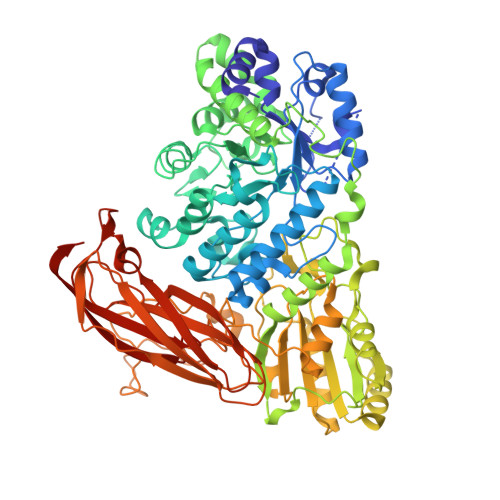
Despite the presence of β-1,2-glucan in nature, few β-1,2-glucan degrading enzymes have been reported to date. Recently, the Lin1839 protein from Listeria innocua was identified as a 1,2-β-oligoglucan phosphorylase. Since the adjacent lin1840 gene in the gene cluster encodes a putative glycoside hydrolase family 3 β-glucosidase, we hypothesized that Lin1840 is also involved in β-1,2-glucan dissimilation. Here we report the functional and structural analysis of Lin1840. A recombinant Lin1840 protein (Lin1840r) showed the highest hydrolytic activity toward sophorose (Glc-β-1,2-Glc) among β-1,2-glucooligosaccharides, suggesting that Lin1840 is a β-glucosidase involved in sophorose degradation. The enzyme also rapidly hydrolyzed laminaribiose (β-1,3), but not cellobiose (β-1,4) or gentiobiose (β-1,6) among β-linked gluco-disaccharides. We determined the crystal structures of Lin1840r in complexes with sophorose and laminaribiose as productive binding forms. In these structures, Arg572 forms many hydrogen bonds with sophorose and laminaribiose at subsite +1, which seems to be a key factor for substrate selectivity. The opposite side of subsite +1 from Arg572 is connected to a large empty space appearing to be subsite +2 for the binding of sophorotriose (Glc-β-1,2-Glc-β-1,2-Glc) in spite of the higher Km value for sophorotriose than that for sophorose. The conformations of sophorose and laminaribiose are almost the same on the Arg572 side but differ on the subsite +2 side that provides no interaction with a substrate. Therefore, Lin1840r is unable to distinguish between sophorose and laminaribiose as substrates. These results provide the first mechanistic insights into β-1,2-glucooligosaccharide recognition by β-glucosidase.
- Department of Applied Biological Science, Faculty of Science and Technology, Tokyo University of Science, Noda, Chiba, Japan.
Organizational Affiliation: