4ZNJ | pdb_00004znj

Thermus Phage P74-26 Large Terminase ATPase domain mutant R139A (I 2 3 space group)


Experimental Data Snapshot

  • Method: X-RAY DIFFRACTION
  • Resolution: 2.53 Å
  • R-Value Free: 
    0.249 (Depositor) 
  • R-Value Work: 
    0.197 (Depositor) 
  • R-Value Observed: 
    0.201 (Depositor) 

Starting Model: experimental
View more details

wwPDB Validation 3D Report Full Report

Validation slider image for 4ZNJ

This is version 1.2 of the entry. See complete history

Literature

Structure and mechanism of the ATPase that powers viral genome packaging.

Hilbert, B.J.Hayes, J.A.Stone, N.P.Duffy, C.M.Sankaran, B.Kelch, B.A.

(2015) Proc Natl Acad Sci U S A 112: E3792-E3799

  • DOI: https://doi.org/10.1073/pnas.1506951112
  • Primary Citation Related Structures: 
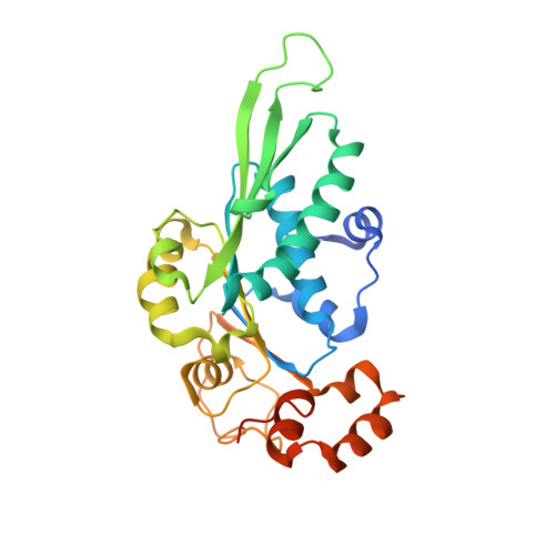
    4ZNI, 4ZNJ, 4ZNK, 4ZNL

  • PubMed Abstract: 

    Many viruses package their genomes into procapsids using an ATPase machine that is among the most powerful known biological motors. However, how this motor couples ATP hydrolysis to DNA translocation is still unknown. Here, we introduce a model system with unique properties for studying motor structure and mechanism. We describe crystal structures of the packaging motor ATPase domain that exhibit nucleotide-dependent conformational changes involving a large rotation of an entire subdomain. We also identify the arginine finger residue that catalyzes ATP hydrolysis in a neighboring motor subunit, illustrating that previous models for motor structure need revision. Our findings allow us to derive a structural model for the motor ring, which we validate using small-angle X-ray scattering and comparisons with previously published data. We illustrate the model's predictive power by identifying the motor's DNA-binding and assembly motifs. Finally, we integrate our results to propose a mechanistic model for DNA translocation by this molecular machine.


  • Organizational Affiliation
    • Department of Biochemistry and Molecular Pharmacology, University of Massachusetts Medical School, Worcester, MA 01605;

Macromolecule Content 

  • Total Structure Weight: 32.07 kDa 
  • Atom Count: 2,126 
  • Modeled Residue Count: 250 
  • Deposited Residue Count: 274 
  • Unique protein chains: 1

Macromolecules

Find similar proteins by:|  3D Structure
Entity ID: 1
MoleculeChains  Sequence LengthOrganismDetailsImage
Phage terminase large subunit274Oshimavirus P7426Mutation(s): 1 
Gene Names: P74p84
EC: 3.6.4 (UniProt), 3.1.21 (UniProt)
UniProt
Find proteins for A7XXR1 (Thermus phage P74-26)
Explore A7XXR1 
Go to UniProtKB:  A7XXR1
Entity Groups
Sequence Clusters30% Identity50% Identity70% Identity90% Identity95% Identity100% Identity
UniProt GroupA7XXR1
Sequence Annotations
Expand
Reference Sequence

Experimental Data & Validation

Experimental Data

  • Method: X-RAY DIFFRACTION
  • Resolution: 2.53 Å
  • R-Value Free:  0.249 (Depositor) 
  • R-Value Work:  0.197 (Depositor) 
  • R-Value Observed: 0.201 (Depositor) 
Space Group: I 2 3
Unit Cell:
Length ( Å )Angle ( ˚ )
a = 129.578α = 90
b = 129.578β = 90
c = 129.578γ = 90
Software Package:
Software NamePurpose
PHENIXrefinement
HKL-3000data reduction
HKL-3000data scaling
PHENIXphasing

Structure Validation

View Full Validation Report


Entry History 

& Funding Information

Deposition Data


Funding OrganizationLocationGrant Number
Hudson Hoagland SocietyUnited States--
Pew Charitable TrustsUnited States--

Revision History  (Full details and data files)

  • Version 1.0: 2015-07-08
    Type: Initial release
  • Version 1.1: 2015-08-05
    Changes: Database references
  • Version 1.2: 2023-09-27
    Changes: Data collection, Database references, Derived calculations, Refinement description